



User Protocol 10:
Immunofluorescent Staining of Adherent Cells in the ibidi µ-Slide 8 Well

Shuntaro Yamada*, Ying Xue, Kamal Mustafa
Centre of Translational Oral Research (TOR)- Tissue Engineering Research Group, University of Bergen
*Corresponding author: Shuntaro.yamada@uib.no
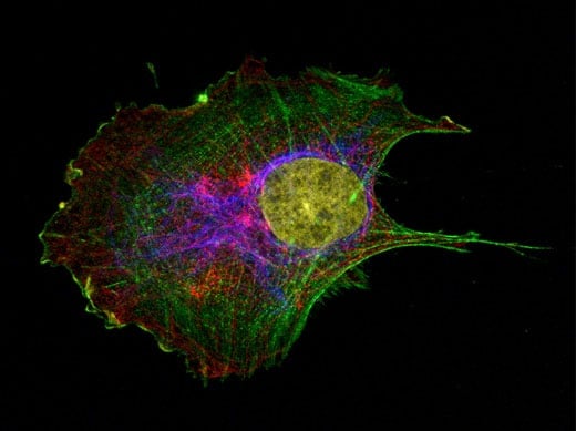
Immunofluorescent staining of adherent cells allows researchers to identify the localization and expression level of proteins of interest, which can also be quantified subsequently. The protocol below guides you to simultaneously label four cellular components (e.g., F-actin, α-Tubulin, stem cell antigen-1, and nuclei) by taking mesenchymal stem cells (MSCs) as an example of adherent cells. The protocol may be adapted to immunofluorescent staining of any adherent cells.
1. Materials and Reagents
Reagents
- Cell culture medium (e.g., α-MEM)
- Fixatives: 4% paraformaldehyde (PFA)
- Tween-20
- Triton X-100
- Phosphate-buffered saline (PBS)
- Normal Goat Serum (NGS)*
- Primary antibody (e.g., Mouse anti-α-Tubulin antibody)
- Secondary antibody (e.g., Goat anti-mouse antibody Alexa FluorTM 555/635)*
- 4',6-diamidino-2-phenylindole (DAPI)
- Phalloidin Alexa FluorTM 488
Note: * The origin of serum should match the origin of the selected secondary antibody (e.g., Normal Goat Serum → goat-derived secondary antibody, Normal Donkey Serum → donkey-derived secondary antibody). Alternatively, 1% BSA can be used.
Equipment
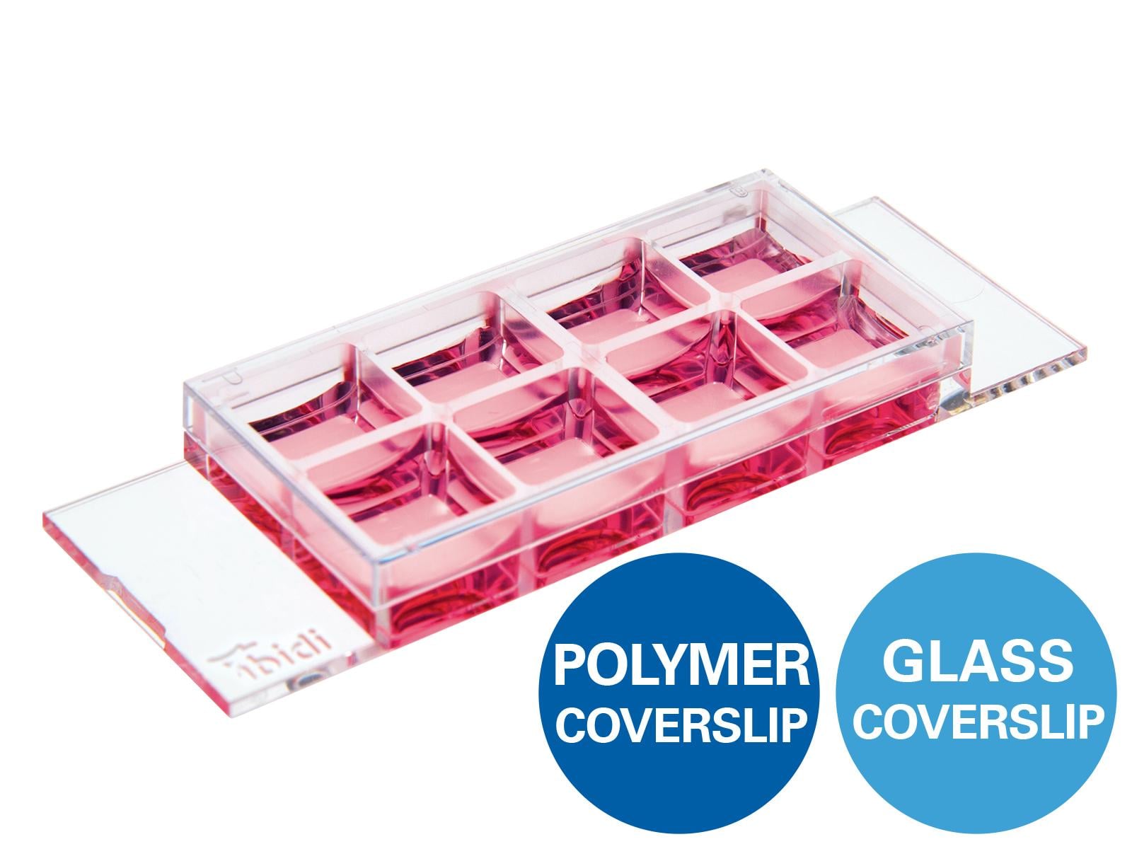
- ibidi µ-Slide 8 Well (Cat. No. 80826; alternatively, the µ-Slide 8 Well high, Cat. No. 80806 can be used).
- Orbital shaker
- Micropipette and tips
- Vortex mixer
- Mini centrifuge
- Fluorescent inverted microscope
Buffer Solutions
- Permeabilization buffer 0.1% Triton X-100 in PBS
- Washing buffer 0.1% Tween-20 in PBS
- Blocking buffer 10% NGS in 0.1% Tween-20 in PBS
- Staining buffer 2% NGS in 0.1% Tween-20 in PBS
| Antibody/stain | Manufacturer | Cat. No. | Working concentration |
|---|---|---|---|
| Primary antibody | |||
| Mouse anti-α-Tubulin antibody | Invitrogen | 62204 | 1:250 |
| Rabbit anti-Sca1/Ly6 antibody | Sigma-Aldrich | AB4336 | 1:500 |
| Secondary antibody | |||
| Goat anti-rabbit antibody Alexa FluorTM 555 | Invitrogen | A32732 | 1:500 |
| Goat anti-mouse antibody Alexa FluorTM 635 | Invitrogen | A31575 | 1:500 |
| Counterstaining | |||
| Phalloidin Alexa FluorTM 488 | Invitrogen | A12379 | 1:250 |
| DAPI | Sigma-Aldrich | D8417 | 0.5 µg/ml |
2. Staining Protocol
2.1 Sample Preparation
- Seed MSCs onto the ibidi µ-Slide 8 Well chambers at a seeding density of 5000–7000/cm2 (Fig. 1)**.
- Allow cells to adhere and grow at 37°C in a 5% CO2 humidified atmosphere until confluency reaches 80%.
Note: ** Seeding density should be optimized according to the type of cells.
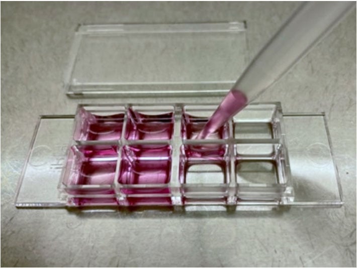
Figure 1. Cell culture using an ibidi µ-Slide 8 Well chamber. For a MSC culture, 5,000–7,000 cells/well are to be seeded into each well. The growth area of each well accounts for 1 cm2, and each well requires 300 µl of cell culture medium.
2.2 Fixation
- Remove the cell culture medium and wash the cells with PBS once.
- Add a sufficient amount of 4% PFA to cover the surface and fix the cells for 15 minutes at room temperature on an orbital shaker.
- Remove the 4% PFA solution and wash the cells with PBS once.
2.3 Permeabilization
- Incubate the cells in the permeabilization buffer (i.e., 0.1% Triton X-100 in PBS) for 10 minutes at room temperature on an orbital shaker.
- Discard the permeabilization buffer and immediately move to the next step.
2.4 Blocking
- Incubate the cells in the blocking buffer (i.e., 10% NGS in 0.1% Tween-20 in PBS) for 60 minutes at room temperature.
- Discard the blocking buffer and immediately move to the next step.
2.5 Primary Antibody Staining
- Prepare the primary staining solution by diluting the primary antibody in the staining buffer (i.e., 2% NGS in 0.1% Tween-20 in PBS). 250–300 μl of staining solution is required for each well.
- Incubate the cells in the primary staining solution overnight at 4°C on an orbital shaker.
- Wash the cells gently in the washing buffer (i.e., 0.1% Tween-20 in PBS) 3–5 times (5 seconds → 5 minutes → 5 minutes → …) on an orbital shaker.
2.6 Secondary Antibody and Counterstaining
- Prepare the secondary staining solution by diluting the secondary antibody, Phalloidin, and DAPI in the staining buffer (Fig. 2)***.
- Incubate the cells in the secondary staining solution for 1 hour at room temperature on an orbital shaker in the dark.
- Wash the cells gently in the washing buffer 3–5 times on an orbital shaker in the dark.
- Keep the samples in the dark until image acquisition.
Note: *** Counterstaining with phalloidin and DAPI can be performed separately. In this case, the cells should be incubated in a staining solution with Phalloidin and DAPI for 20 minutes at room temperature in the dark after the secondary antibody staining.
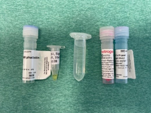
Figure 2. Preparation of secondary antibody and counterstaining solution. For 8 wells of the ibidi µ-Slide 8 Well, 2 ml of staining solution are required (i.e., 250 µl per well). The secondary antibody (1:500), Phalloidin (1:250) and DAPI (0.5 µg/ml) can be simultaneously added in the staining buffer for secondary antibody incubation and counterstaining.
3. Image Acquisition
| Fluorochromes | Excitation max | Emission max |
|---|---|---|
| Alexa FluorTM 488 | 490 | 525 |
| Alexa FluorTM 555 | 558 | 573 |
| Alexa FluorTM 635 | 633 | 647 |
| DAPI | 350 | 470 |
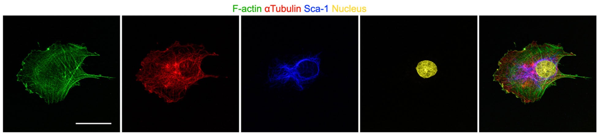
Figure 3. A mesenchymal stem cell was treated with 1 nM Narciclasin to activate Rho-ROCK signaling. The image was acquired with a Leica SP8 Confocal Microscope with 93x oil immersion lens using a sequential scan mode. F-actin, α-Tubulin, Sca-1, and nuclei are depicted in green, red, blue, and yellow, respectively. Scale bar 50 µm.
4. General Tips
- Prepare a fresh 4% PFA solution prior to fixation. 4% PFA can be stored at -20°C for less than 3 months.
- Do not dry the cells during the entire process.
- It is recommended to optimize the working concentration of antibody. The reactivity may vary due to lot-to-lot variability.
- Before adding antibodies to the staining buffer, briefly centrifuge and use only the supernatant. The antibody may be precipitated.
- Phalloidin staining is not compatible with alcohol-based (e.g., ice-cold methanol) or acid-based (e.g., trichloroacetic acid) fixation and is not compatible with most enzymatic or heat-induced epitope retrieval processes.
This User Protocol is an ibidi peer-reviewed protocol from an actual user. ibidi does not guarantee its functionality or reproducibility. For this User Protocol, ibidi provides only limited support. Please contact the corresponding author for detailed information.
For research use only.