



User Protocol 15:
Protocol for Generating Self-Organized Tubular Bile Duct Organoids from hiPSC-Derived Hepatobiliary Cells on the µ-Pattern ibiTreat
Kim Whye Leong*, Samuel Wang, Hanry Yu
Mechanobiology Institute, National University of Singapore
*Corresponding author, e-mail: kimwhyeleong@u.nus.edu
The intrahepatic bile duct makes up around 5% of the liver mass and is difficult to isolate from human biopsy samples. This is coupled with difficulty in propagating the primary cholangiocytes, limiting the establishment of in vitro models of the intrahepatic bile duct for studying biliary disorders and advancing drug discovery. To circumvent this, protocols to differentiate human induced pluripotent stem cells (hiPSCs) into functional cholangiocytes have been developed.
In this protocol, we show the differentiation of hiPSCs into hepatobiliary cells and induce tubular organoid formation using a collagen I sandwich configuration. By incorporating the use of ibidi µ-Pattern, this protocol provides a possible drug screening platform with the tubular bile duct organoids.
1. Related Documents
2. Materials and Reagents
- hiPSC cell lines from Allen Institute for Cell Science:
AICS-0023 (AICS23, mEGFP-TJP1), AICS-0024 (AICS24, mEGFP-MYH10), or AICS-0058-067 (AICS58, mEGFP-CTNNB1)
Note: All cell lines can be used for differentiation, the different tagged proteins visualize different cell parameters.
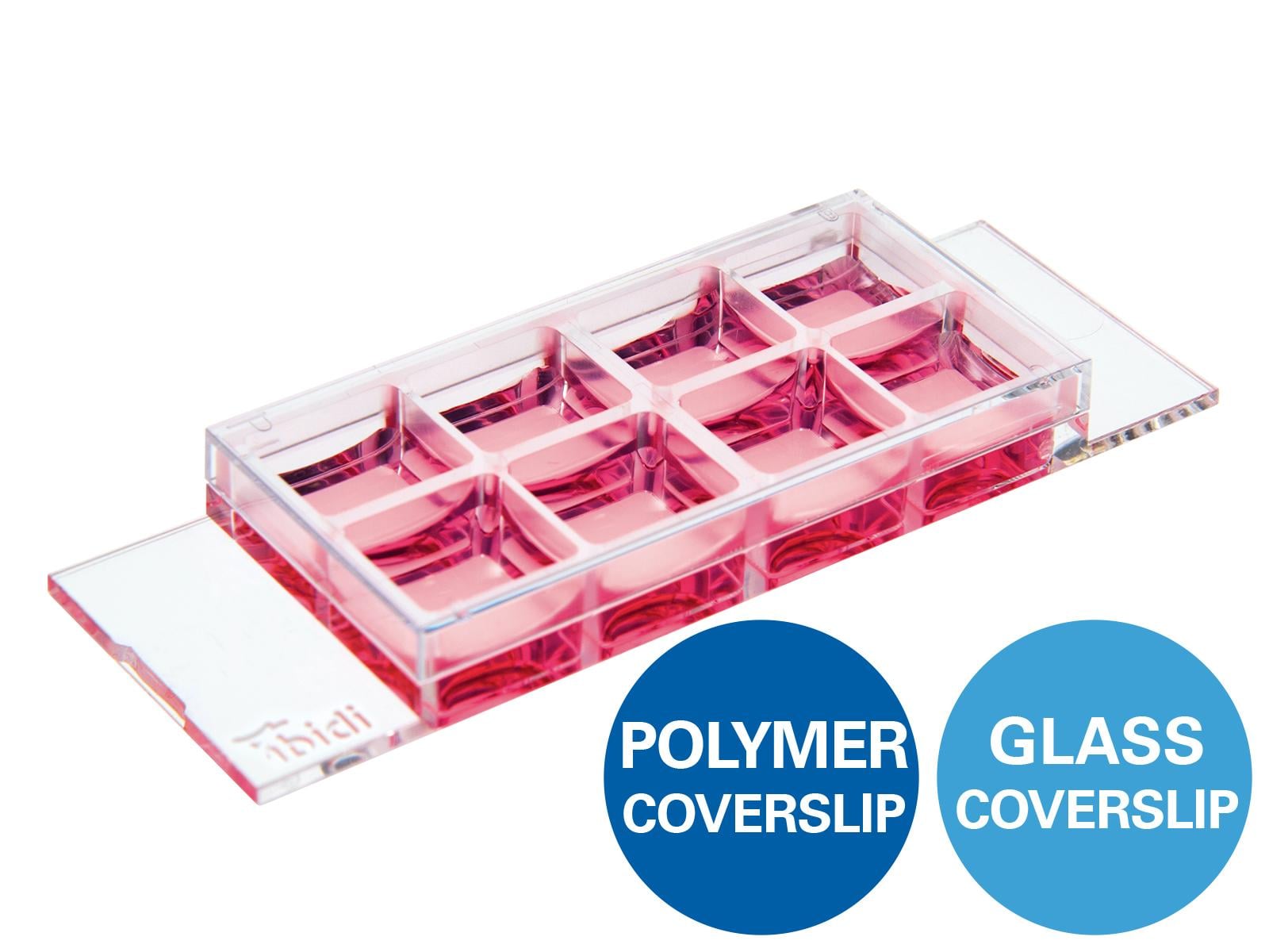
- µ-Slide 8 Well high µ-Pattern ibiTreat, cir500, pit1000, hex (ibidi, 83813)
- 12 well plate (Thermo Scientific™, 150628)
- Bovine collagen I, prepared as specified by the manufacturer (PureCol®, 5005)
- 0.01 N HCl
- 0.1 N NaOH
- 10× DPBS (Gibco™, 14080055)
- 1× DPBS (Gibco™, 14040133)
- 1× PBS
- Trypsin-EDTA (0.5%), no phenol red (Gibco™, 15400054)
- mTeSR™ Plus (STEMCELL Technologies, 100-0276)
- Growth factor-reduced (GFR) Matrigel® (Corning, 354230)
- Y-27632 (Sigma Aldrich, Y0503)
The following differentiation media were modified from previous protocols used to differentiate hepatocytes [1], cholangiocytes [2] or both [3,4].
RPMI medium
To 500 ml of RPMI-1640 (Gibco™, 72400047), add:
- 10 ml B-27™ supplement without insulin (Gibco™, A1895601)
- 5 ml MEM non-essential amino acids solution (Gibco™, 11140050)
- 5 ml pen/strep (Gibco™, 15140122)
Definitive Endoderm (DE) medium
To the prepared RPMI medium, add:
- 100 ng/ml Activin A (R&D Systems, 338-AC)
- 20 ng/ml bFGF (R&D Systems, 233-FB),
- 10 ng/ml BMP4 (R&D Systems, 314-BP)
Foregut Progenitor (FP) medium
To the prepared RPMI medium, add:
- 50 ng/ml Activin A (R&D Systems, 338-AC)
Hepatoblast (HB) medium
To the prepared RPMI medium, add:
- 0.3 µl/ml insulin (Gibco™, 12585014)
- 10 µM SB 431542 (STEMCELL Technologies, 72232_C)
- 50 ng/ml BMP4 (R&D Systems, 314-BP)
Quench medium
To the prepared RPMI medium, add:
- 10% FBS (HyClone, SH30070.01)
Advanced DMEM/F12 medium
To 500 ml of advanced DMEM/F12 medium (Gibco™, 12634028), add:
- 10 ml B-27™ supplement (Gibco™, 17504044)
- 5 ml N2 (Gibco™, 17502048)
- 5 ml HEPES (Gibco™, 15630106)
- 10 mM nicotinamide (Sigma-Aldrich, N3376)
- 50 nM A83-01 (STEMCELL Technologies, 72022)
- 0.5 µM dbCAMP (STEMCELL Technologies, 73882)
- 100 ng/ml EGF (R&D Systems, 236-EG)
- 20 ng/ml HGF (R&D Systems, 294-HG-005/CF)
- 500 ng/ml R-spondin
- 50 ng/ml FGF10 (R&D Systems, 1277-JG-050)
- 5 ml pen/strep (Gibco™, 15140122)
Liver Progenitor (LP) medium
To the prepared advanced DMEM/F12 medium, add:
- 2 µg/ml Jagged-1 (Anaspec, AS-61298)
- 3 µM retinoic acid (Sigma-Aldrich, R2625)
Hepatobiliary (HepBili) medium
To the prepared advanced DMEM/F12 medium, add:
- 30 µM dexamethasone (STEMCELL Technologies, 72092)
- 20 ng/ml BMP4 (R&D Systems, 314-BP)
3. Procedure
3.1 Cell Seeding
- Passage and maintain hiPSCs according to protocol version 1.7 2020 from Allen Institute for Cell Science [5].
- To prepare for differentiation, passage the cells and seed at 3 × 10⁵ cells/cm2 in mTeSR™ Plus on a GFR Matrigel-coated 12 well plate according to the lot-specified dilution factor.
3.2 Differentiation of hiPSCs into HB Cells (Day 1–9)
- Day 1–2: Ensure that the hiPSCs are confluent and attached after seeding. Aspirate the mTeSR™ Plus and add the DE medium. Change medium every 24 hours.
- Day 3–5: Replace the DE medium with FP medium. Change medium every 24 hours for three days.
- Day 6–9: Replace the FP medium with HB medium. Change medium every 24 hours for four days.
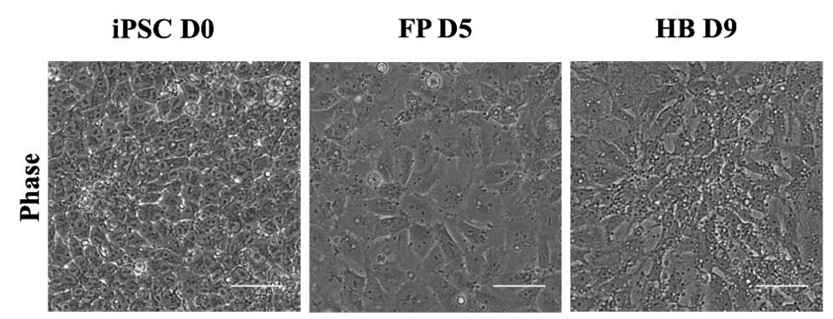
Figure 1. Differentiation of hiPSCs into HB cells. Phase contrast images of human induced pluripotent stem cell (iPSC, Day 0), foregut progenitor (FP, end of Day 5), and hepatoblast (HB, beginning of Day 9) stages during differentiation. Scale bars, 20 µm.
3.3 Seeding Hepatoblast Cells on the ibidi µ-Pattern
- Coat the µ-Slide 8 Well high µ-Pattern ibiTreat with 1.5 mg/ml bovine collagen I at 150 µl per well for 30 minutes.
- Wash the µ-Slides with 1× PBS five times.
- Aspirate the HB medium from day 9 from the 12 well plate and wash with 1× PBS twice.
- Add 300 µl of trypsin-EDTA to the cells in each well of the 12 well plate and incubate at 37°C for 3 minutes to detach the cells.
- Transfer the cells into a 15 ml Falcon tube and add quench medium in a 1:1 ratio with respect to trypsin volume.
- Spin down at 180 g for 5 minutes.
- Resuspend the cell pellet with HB medium supplemented with 10 µM of Y-27632 and count the cells.
- Seed the cells into the µ-Slides with a density of 1 × 10⁵ cells/cm2 with a minimum volume of 300 µl per well.
- Incubate the cells overnight at 37°C, 5% CO2 and humidified atmosphere.
3.4 Differentiation to Hepatobiliary Cells (Day 10–23)
- Day 10–13: Aspirate HB medium with Y-27632 from the µ-Slide and replace it with LP medium. Change the medium every 24 hours for another three days.
- Day 14–23: Replace the LP medium with HepBili medium. Change the medium every 2–3 days for 10 days.
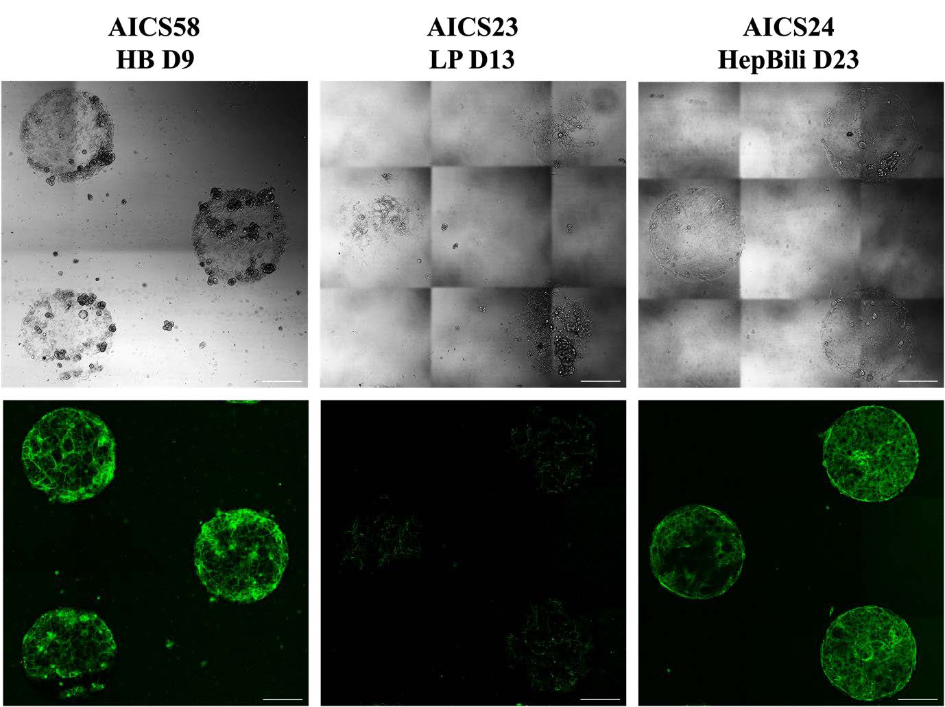
Figure 2. Differentiation to hepatobiliary cells on the µ-Pattern ibiTreat. Phase contrast and fluorescence images of hepatoblast (HB, end of Day 9), liver progenitor (LP, end of Day 13), and hepatobiliary (HepBili, end of Day 23) stages on a 500 µm diameter micropattern. AICS58 HB express mEGFP-tagged CTNNB1 marking the cell-cell boundaries, useful for cell segmentation. AICS23 express mEGFP-tagged TJP1 marking the tight junction near the apical membrane to inform on cell polarity. AICS24 express mEGFP-tagged MYH10 marking myosin IIB, which can be used to infer cell-dependent contractility. Scale bars, 200 µm.
3.5 Inducing Tubular Organoid Formation
- Prepare bovine collagen I gel according to Table 1 for the necessary volume.
- Apply a minimum of 300 µl collagen I gel to each well of the µ-Slide at 150 µl/cm2 and incubate the cells overnight at 37°C, 5% CO2 and humidified atmosphere.
- Add HepBili medium to the culture the next day.
Table 1. Reagents for collagen I gel mixture.
| Order of addition | Reagent | Ratio |
|---|---|---|
| 1 | HepBili medium | 32 |
| 2 | 1× DPBS | 6 |
| 3 | 10× DPBS | 1 |
| 4 | NaOH (0.1 N) | 1 |
| 5 | Bovine Collagen I | 8 |
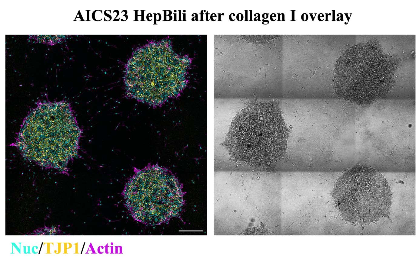
Figure 3. Induction of tubular organoids on micropattern 24 hours after collagen I overlay. Phase contrast and composite fluorescence image of maximum intensity projected nuclei (DAPI), actin (Phalloidin) and mEGFP-tagged TJP1. After overlaying with collagen, the cells begin to migrate into the gel and form tubular structures. TJP1 is localized near the apical membrane, demarcating the lumen formed by the hepatobiliary cells. Scale bar, 200 µm.
4. References
- Cai, J., Fisher, J., Urick, A., Wagner, T., Cayo, K. T. M., Nagaoka, M., & Duncan, S. A. (2015). Protocol for directed differentiation of human pluripotent stem cells toward a hepatocyte fate. African Scientist, 16(1), 13-26.
- Sampaziotis, F., de Brito, M. C., Geti, I., Bertero, A., Hannan, N. R., & Vallier, L. (2017). Directed differentiation of human induced pluripotent stem cells into functional cholangiocyte-like cells. Nature protocols, 12(4), 814-827.
- Huch, M., Dorrell, C., Boj, S. F., Van Es, J. H., Li, V. S., Van De Wetering, M., ... & Clevers, H. (2013). In vitro expansion of single Lgr5+ liver stem cells induced by Wnt-driven regeneration. Nature, 494(7436), 247-250.
- Ramli, M. N. B., Lim, Y. S., Koe, C. T., Demircioglu, D., Tng, W., Gonzales, K. A. U., ... & Chan, Y. S. (2020). Human pluripotent stem cell-derived organoids as models of liver disease. Gastroenterology, 159(4), 1471-1486.
- Allen Institute for Cell Science. (2020). Culture and Freezing Methods for WTC Derived AICS hiPSC Lines. https://www.allencell.org/uploads/8/1/9/9/81996008/aics_wtc_culture_sop_external_v1-7_200211.pdf
This User Protocol is an ibidi peer-reviewed protocol from an actual user. ibidi does not guarantee its functionality or reproducibility. For this User Protocol, ibidi provides only limited support. Please contact the corresponding author for detailed information.
For research use only.