



User Protocol 08:
Protocol for an Adhesion Assay Using Cell Culture Under Unidirectional Flow
Hannah Manz, Andreas Brandl*
Department of Internal Medicine II, Würzburg University Hospital, Würzburg, Germany.
*Corresponding author. E-mail address: brandl_a@ukw.dev
This protocol describes an adhesion assay under flow that was established to investigate the role of specific cellular surface molecules, in order to determine their potential interaction during cell attachment and adhesion to other cell types.
For our approach, we used pre-stained murine tumor cells (multiple myeloma: MM) and murine endothelial cells (EC), then blocked one of our molecules of interest using a monoclonal antibody. Briefly, ECs were seeded into ibidi channel µ-Slides I 0.6 mm Luer and cultured under constant flow for 24 h. To start the co-culture, labeled MM cells were added to the flow reservoirs and the flow was continued. 24 h later, the setups were either treated with antibody (to block the molecule of interest) or the respective isotype control, and were kept under flow for 24 h. The next day, the µ-Slides were disconnected from the Fluidic Units (FU) and washed to remove non-adherent MM cells. To analyze the adherence of the labeled MM cells to the ECs, fluorescent images of the µ-channels were acquired using confocal microscopy.
1. Materials and Reagents
- MOPC multiple myeloma (MM) cell line (ATCC, TIB-23™)
- EOMA endothelial cell (EC) line (ATCC, CRL-2586™)
- RPMI culture medium with 10% FCS (Fisher Scientific, 11530586)
- Endothelial Cell Growth Medium (EC medium, CellBiologics, M1168)
- PBS (PAN-Biotech, P04-361000)
- Cell dissociation solution non-enzymatic (Sigma-Aldrich, C5914-100ML)
- Trypan blue solution (Sigma-Aldrich, 93595)
- CellTracker™ Green CMFDA Dye (Thermo Fisher, C7025)
2. Equipment and Settings
- ibidi Pump System with 2 Fluidic Units (FU), each with holders for 2 ml reservoirs (ibidi, 10977)
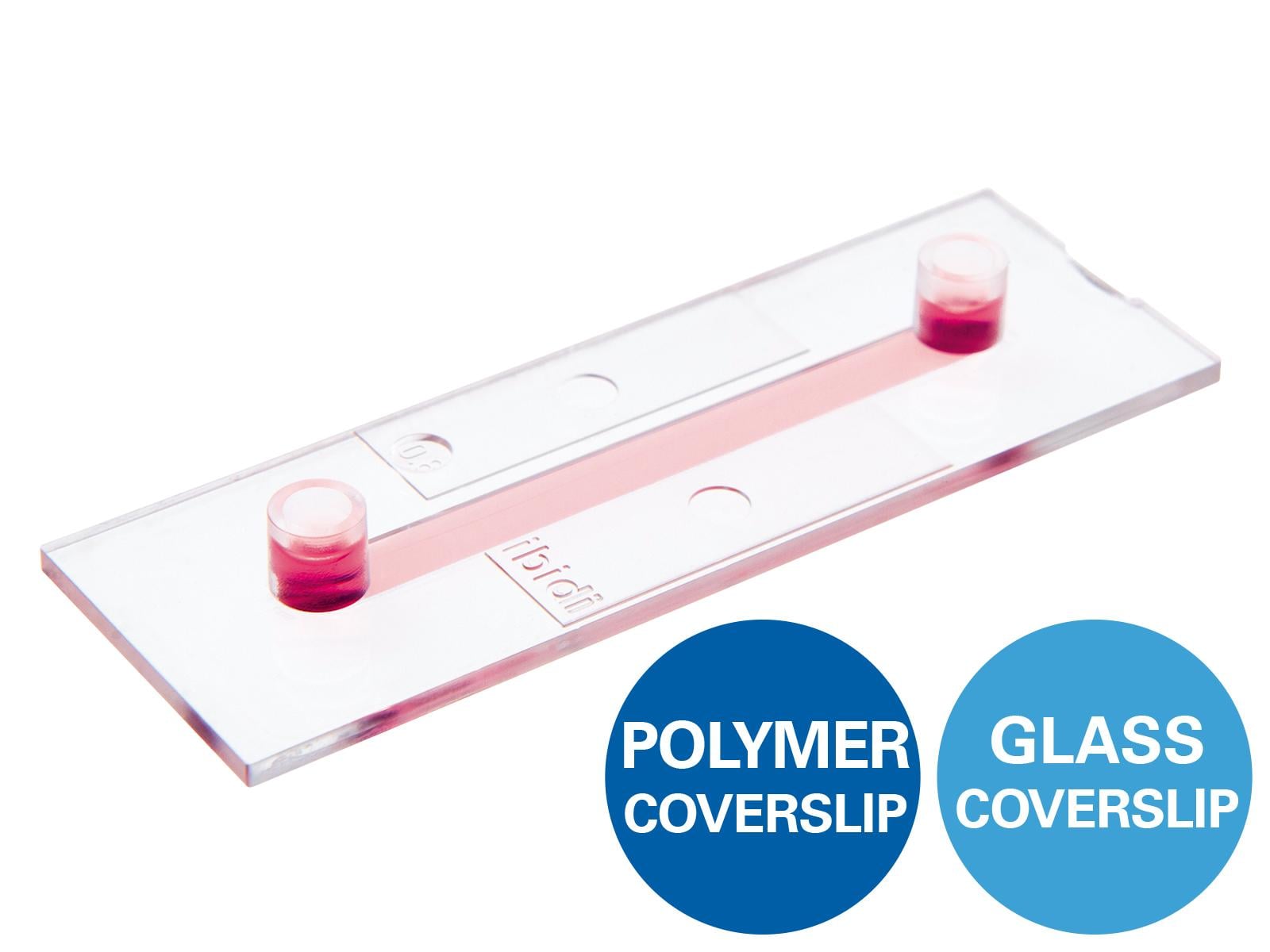
- ibidi µ-Slide I 0.6 mm Luer, ibiTreat (ibidi, 80186)
- ibidi Perfusion Set BLUE, 15 cm, ID 0.8 mm (ibidi, 10961)
- ibidi Filter/Reservoir Set, 2 ml (ibidi, 10972)
- Laminar flow hood with cell culture equipment
- Incubator (all incubation steps were carried out at 37°C and 5% CO2)
- Fluorescence microscope with appropriate filter sets and camera
- Viscosity: 0.0072 (dyn x s)/cm²
3. Procedure
- Prepare EOMA cell dilution: Detach the previously cultured EOMA cells using non-enzymatic cell dissociation solution according to the manufacturer’s protocol. Count the cells using a hemocytometer (Neubauer chamber). Dilute the cells to a concentration of 1.2 x 10⁶ cells/ml in EC medium.
- Seed EOMA cells: Fill 3 µ-Slides I 0.6 mm Luer with 150 µl of the EOMA cell dilution per slide (1.75 x 10⁵ EOMA cells per µ-Slide), then incubate them for 3 h.
Table 1. Experimental setup.
| µ-Slide No. | Flow rate [ml/min] | Shear stress [dyn/cm²] | Pressure [mbar] | Unidirectional switching time [s] | Cycle duration [s] |
|---|---|---|---|---|---|
| #1 (isotype ctrl) | 1.15 | 0.5 | 8.2 | 100 | infinite |
| #2 (antibody) | 1.15 | 0.5 | 8.2 | 100 | infinite |
| #3 (static control) | 0 | 0 | n/a | n/a | n/a |
- Start flow: Three hours after seeding, connect 2 µ-Slides to the FUs using a total amount of 2.7 ml EC medium. Set the shear stress on the EOMA cell surface to 0.5 dyn/cm² using a pressure of 8.2 mbar. Measure the flow rate and use the mean from both FUs to calculate the calibration factor. Incubate the FUs and the connected slides overnight. The third µ-Slide with EOMA cells serves as a static control. Incubate it overnight without any flow.
- Prepare MM cells: Stain 6 x 10⁵ MM cells in 600 µl RPMI medium (w/o FCS) with CellTracker Green (CMFDA) dye according to the manufacturer’s protocol. After labeling, centrifuge the cells and count them using a hemocytometer.
- Start co-culture with MM cells: Stop the flow and remove the EC medium from the FU reservoirs under sterile conditions. To start the co-culture, mix RPMI medium (with 10% FCS) and EC medium in a 1:1 proportion and fill a total volume of 2.5 ml of this mixture containing 1.5 x 10⁵ MM cells equally into both FU reservoirs. Reconnect the FUs to the pump system and continue the flow for 24 h.
- Molecule blocking: 24 h after the addition of the MM cells to the ECs, treat the cells with 100 µg/ml antibody (Slide #2) or the corresponding isotype control (Slide #1), by adding them directly into the particular reservoirs of the FUs without replacing the medium. Keep the incubation under flow for another 24 h.
- Washing and imaging: Disconnect the µ-Slides from the FUs. Wash the cells once with PBS to remove any non-adherent cells. Acquire fluorescent images using confocal microscopy to visualize labeled MM cells that are attached to the EC layer.
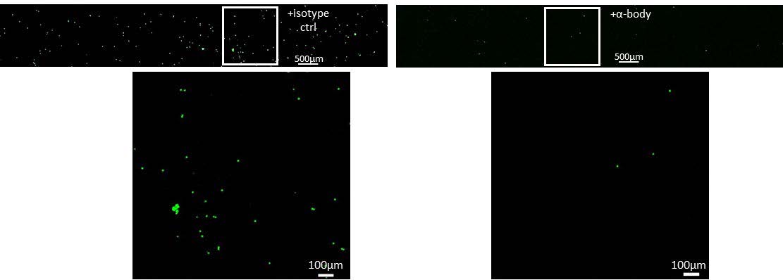
Figure 1. MM cell adherence to ECs is reduced when blocking a specific surface molecule (right panel) compared to isotype control treatment (left panel).
This User Protocol is an ibidi peer-reviewed protocol from an actual user. ibidi does not guarantee its functionality or reproducibility. For this User Protocol, ibidi provides only limited support. Please contact the corresponding author for detailed information.
For research use only.