



User Protocol 12:
Time-Lapse Microscopy of Zebrafish Larvae in the ibidi µ-Slide 2 Well Co-Culture
Friederike Kessel*
Neural Development and Regeneration, Center for Regenerative Therapies Dresden (CRTD), Technical University Dresden, Dresden, Germany
*Corresponding author. E-mail address: friederike.kessel@tu-dresden.de
This User Protocol describes the process of embedding zebrafish larvae in low percentage low melting point agarose for time-lapse microscopy. Depending on the scientific question, different stages of development can be observed in the agarose-embedded larvae: This can either include the initial 48 hours of development in the chorion, or the time after hatching (48–120 hours post fertilization).
1. Related Documents
2. Materials and Reagents
Animals
- Zebrafish larvae (up to 120 hours post fertilization)
Buffers and Solutions
Fish Water (60x)
- 172 g NaCl (ROTH, P029.2)
- 7.6 g KCl (ROTH, 6781.1)
- 29 g CaCl2 x 2 H2O (ROTH, CN92.2)
- 49 g MgSO4 x 7 H2O (ROTH, P027.1)
- Ad 10 l with deionized water
Fish Water (1x)
- 160 ml fish water (60x)
- 20 ml 0.01% (w/v) Methylene Blue (Sigma-Aldrich, M9140) in aqua dest
- Ad 10 l with deionized water
Tricaine Stock Solution
- 4 g/l Tricaine/MS-222 (Sigma Aldrich, A5040)
- 0.02 M Tris (ROTH, 4855.2), pH 9
- In aqua dest
Tricaine Use Solution
- Dilute Tricaine Stock Solution in Fish Water (1x) to a final concentration of 160 mg/l (1:25)
0.5% Low Melting Point Agarose (LMPA)
- Can be prepared in bulk and split into small aliquots for each experiment
- Boil 0.5% (w/v) Low Melting Point Agarose (Biozym, 840101) in fish water (1x) in a microwave until agarose is completely dissolved
- Let the agarose cool down (~35°C)
- Add tricaine stock solution to a final concentration of 80 mg/l (1:50)
The final concentration of LMPA can be adjusted. For example, we successfully used up to 2% LMPA but decided to execute our experiments with 0.5% LMPA because it is less likely to influence physiological processes.
Equipment
- Thermal mixer (e.g., Eppendorf, ThermoMixer C)
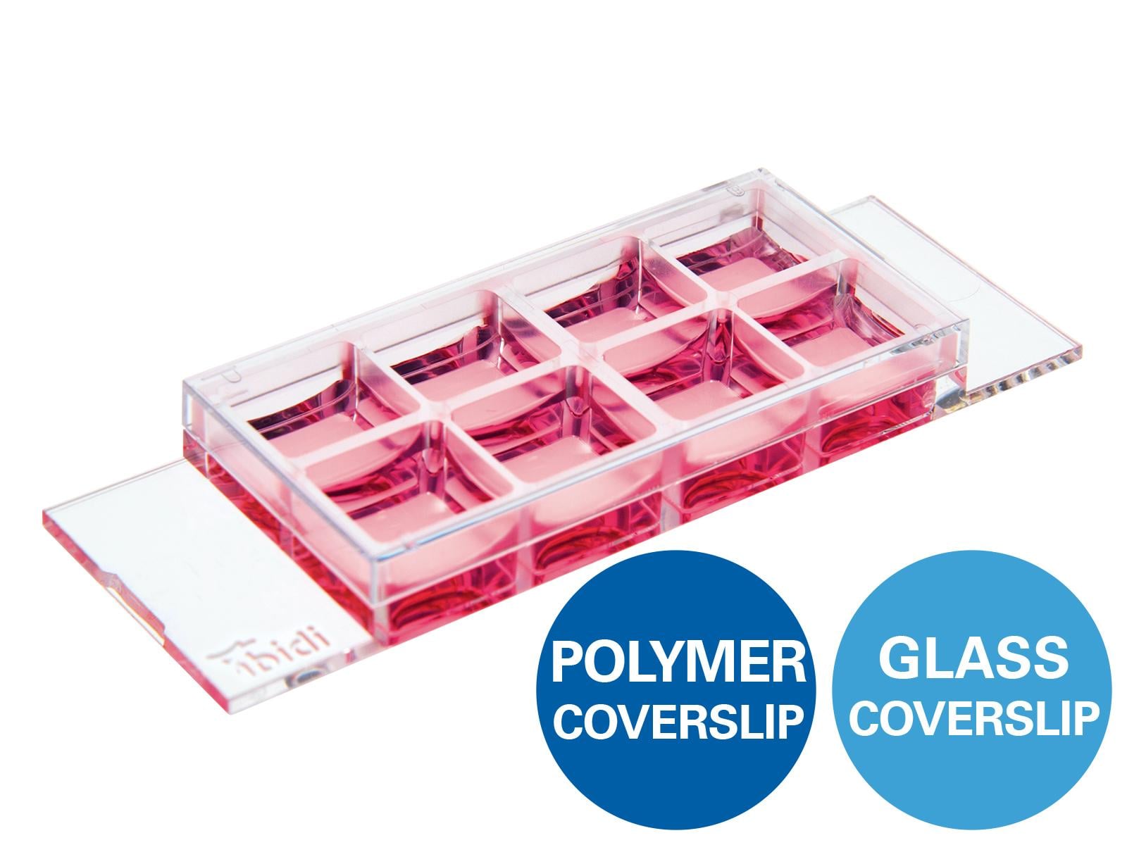
- µ-Slide 2 Well Co-Culture, ibiTreat (ibidi, 81806)
- Plastic transfer pipettes
- Optional: small brush
- Microscope
3. Procedure
Preparations
Before starting the experiment, prepare the LMPA as described in the "Buffers and Solutions" Section. Keep at 35°C until needed. If the agarose has been prepared in advance, reheat the aliquot in a thermal mixer (~80°C) and let the agarose cool down (~35°C).
Mounting
Please read the Instructions before working with the µ-Slide 2 Well Co-Culture.
In some experiments in our group, mechanical lesions were induced to the zebrafish spinal cord. If doing this, it is recommended to let the larvae recover for at least 30 min prior to mounting. After transection of the spinal cord, the initial survival after embedding in agarose is 80%. All these larvae also survive the observation period of 48 h in agarose. The initial survival of uninjured larvae is close to 100%.
- Anesthetize the zebrafish larvae in Tricaine Use Solution for 2 minutes.
- Place one larva into each µ-Slide 2 Well Co-Culture chamber with a bit of excess Fish Water using a plastic transfer pipette.
- Perform the following steps for each larva separately (to avoid drying out):
- Remove all Fish Water with a plastic transfer pipette.
- Add 50 µl of LMPA into the minor well to cover the larva.
- Using a brush or small pipette tip, orient the larva to the proper position (a lateral position, as horizontal and straight as possible with a uniform alignment, i.e., always facing left with the yolk at the bottom).
- Let the LMPA gel solidify (0.5% gel ~20–30 min).
- Optional: cover the solidified LMPA gel with 600 µl 1x Fish Water containing 80 mg/l tricaine stock solution for continuous anesthesia during imaging.
- Cover the µ-Slide 2 Well Co-Culture with its lid to avoid evaporation.
- The mounted zebrafish larvae are now ready for imaging.
Adding Fish Water on top of the agarose is optional since the specimens do not dry out during the 48 h of imaging if the lid is tightly closed. This also enables upright microscopy without risking spills.
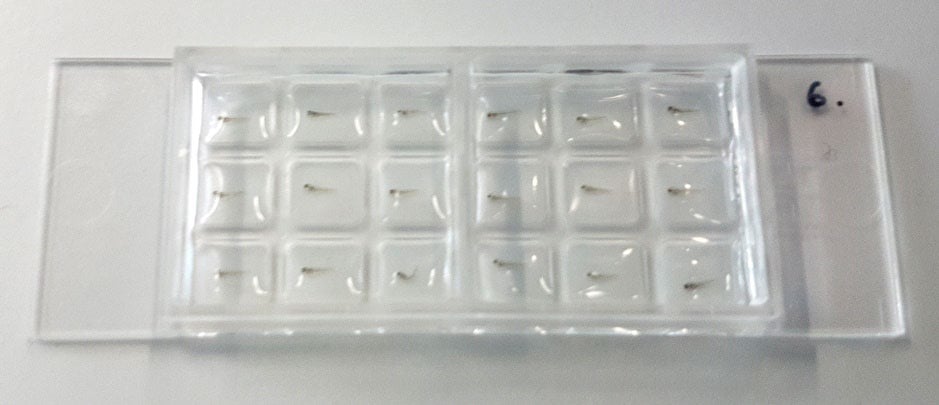
Figure 1. Zebrafish larvae embedded in LMPA in a µ-Slide 2 Well Co-Culture.
Time-lapse Microscopy
This procedure for sample preparation has been used for continuous time-lapse microscopy as well as for snapshots of certain fields of view at different time points with various microscopes of the Light Microscopy Facility, a core facility of CRTD at Technical University Dresden. This included inverted and upright, widefield and confocal microscopes.
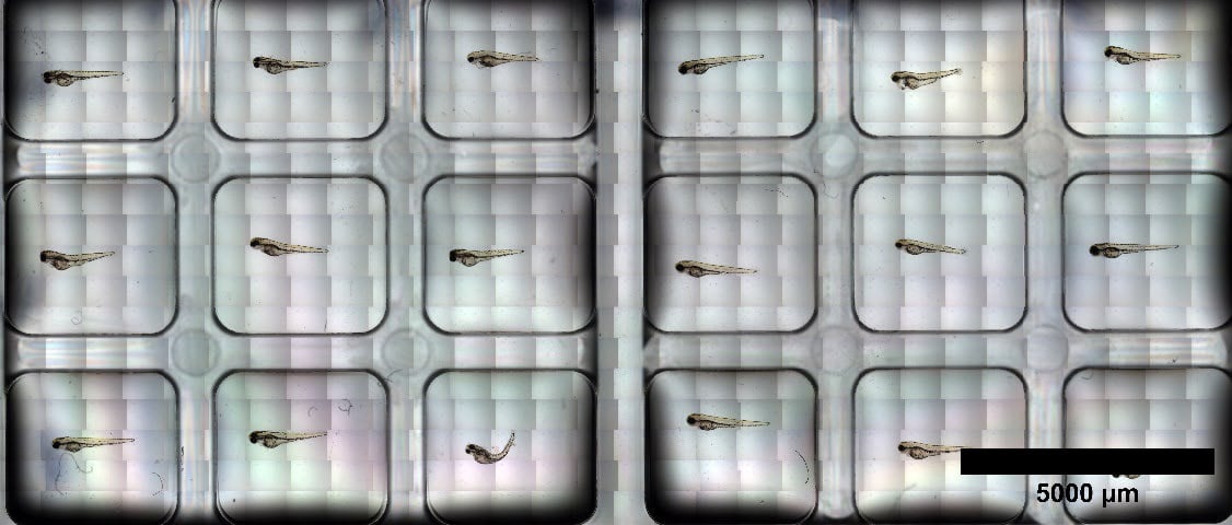
Figure 2. A) Brightfield tile-scan of the entire µ-Slide 2 Well Co-Culture with a total of 18 zebrafish larvae 72 hours post fertilization, 3 hours after embedding in agarose.
Movie: The attached example shows the Z-projection of a short 20 minute 3D-time-lapse, 24 hours post mechanical transection of the spinal cord in a transgenic mpeg1:mCherry zebrafish larva 3 days post fertilization. The fluorescently labeled cells are macrophages and microglia. This time-lapse was acquired with the Dragonfly Spinning Disk microscope (Andor). Watch the movie here.
This User Protocol is an ibidi peer-reviewed protocol from an actual user. ibidi does not guarantee its functionality or reproducibility. For this User Protocol, ibidi provides only limited support. Please contact the corresponding author for detailed information.
For research use only.