




MV 75: Fluorescence Staining in the µ-Slide VI
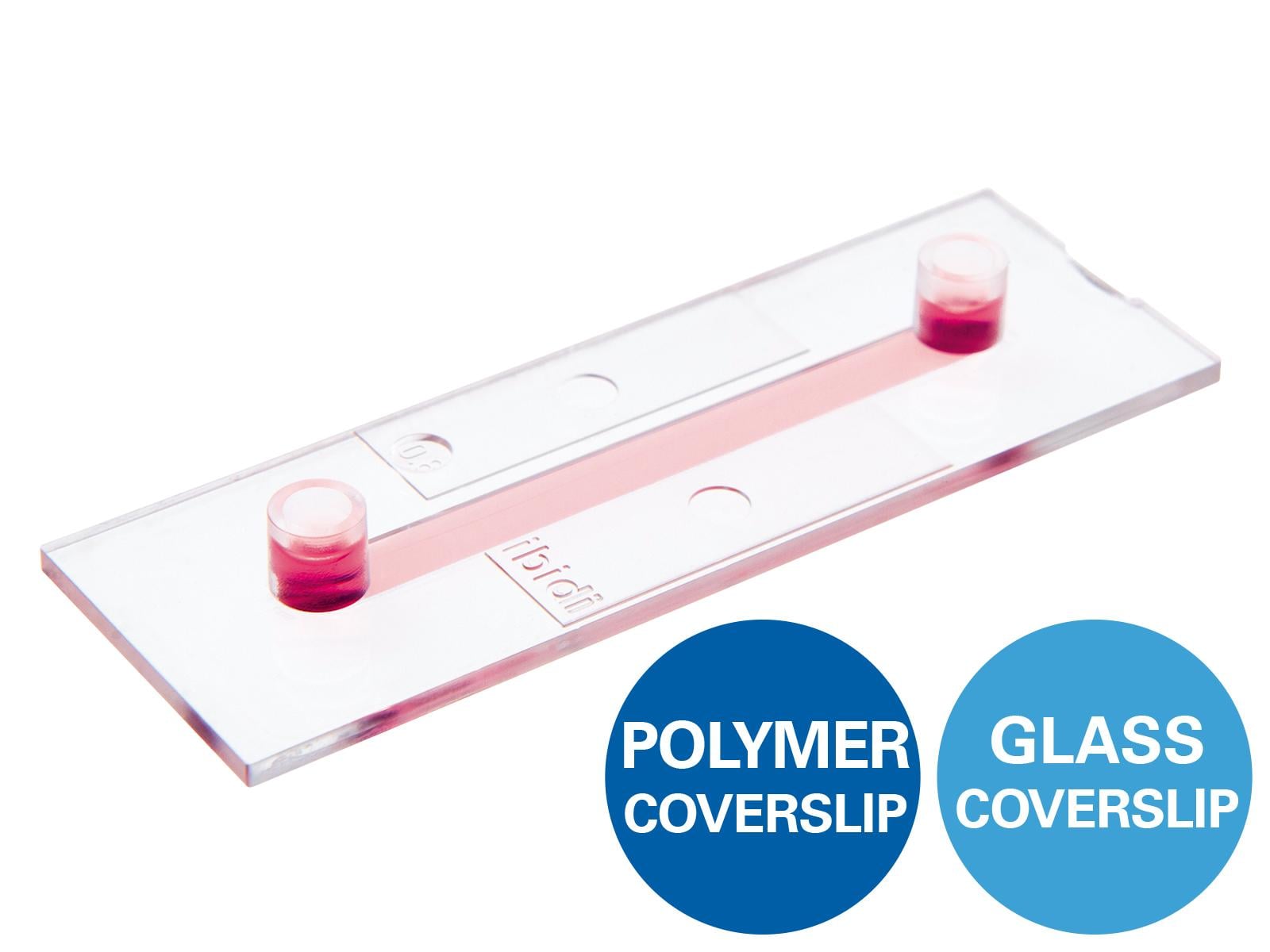
In this video, we demonstrate an immunofluorescence protocol using the ibidi µ-Slide VI 0.4. For detailed step-by-step instructions, please refer to Application Note 09: Fluorescence Staining Using a µ-Slide VI (PDF).
This all-in-one chamber simplifies immunofluorescence protocols by combining cell culture, staining, and imaging in one slide—using only small numbers of cells and minimal reagent volumes.
Key Features of the µ-Slide VI 0.4
- Six parallel channels for efficient, comparative experiments
- Homogeneous cell distribution over the channel surface
- Only 30 µl per channel—cost-effective with reduced reagent consumption
- Compatible with high-resolution and super-resolution microscopy
- Suitable for static or flow conditions with defined shear stress
Applications
- Immunofluorescence assays
- Live cell imaging under static or flow conditions
- Parallel screenings using multichannel pipettes
- High-resolution and super-resolution fluorescence microscopy (e.g., STED, confocal)
Detailed product information: µ-Slide VI , AN 09: Fluorescence Staining Using a µ-Slide VI (PDF)