


Cells Under Shear Stress: Wall Shear Stress, Flow Types, and Applications
Cells under shear stress are exposed to mechanical forces generated by fluid flow along the cell surface. In cell-based assays, wall shear stress is especially relevant for adherent cells such as endothelial and epithelial cells, where it can influence morphology, alignment, adhesion, barrier function, gene expression, and mechanotransduction. This page focuses on wall shear stress. For applications involving continuous medium exchange, nutrient supply, and low-shear perfusion, see Perfusion-Based Cell Culture..
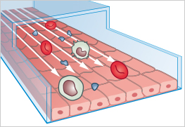
Many adherent cell types are naturally surrounded by moving fluids. Examples include vascular endothelial cells that form the inner layer of blood vessels, lymphatic endothelial cells that form the inner layer of lymphatic vessels, and epithelial cells in organs such as the kidney and lung.
The movement of fluid along the cell surface generates wall shear stress, a mechanical force that can influence cell morphology, alignment, adhesion, barrier function, signaling, and gene expression.
In standard static in vitro culture, this mechanical stimulus is missing. Shear stress assays help reproduce a key aspect of the in vivo microenvironment and can therefore provide more physiologically relevant cell behavior than static culture conditions.
ibidi Blog Articles |
Are you curious about 3 Reasons Why You Should Cultivate Endothelial Cells Under Flow? Get a concise overview in the ibidi Blog.
Discover how cell culture under flow is used to simulate endothelial dysfunction in our blog article ”Shear Genius: Microfluidics in Atherosclerosis Research”.
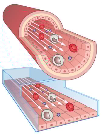
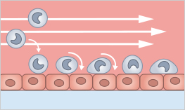
Endothelial cells in blood vessels (top) are under continuous flow. Using in vitro cell culture under flow (bottom), these physiological conditions can be simulated.
Wall shear stress is the mechanical force induced by the friction of liquid against the apical cell membrane. In vivo, several adherent cell types are exposed to mechanical shear stress in biofluidic systems, such as blood and lymphatic vessels or nephrons.
This mechanical stimulus can influence several cellular responses, including:
- cell morphology and alignment,
- cytoskeletal organization,
- ion channel activation and mechanotransduction,
- gene expression and inflammatory signaling,
- adhesion properties and barrier function,
- organization of the whole cell layer.

Wall shear stress is commonly measured in dyne/cm² (dyn/cm²). Physiological wall shear stress varies strongly between vessel types, tissues, and organisms, ranging from 0.5 to 120 dyn/cm² and depend on the vessel type (e.g., artery or vein), the tissue (e.g., brain, connective tissue, or heart), and the size of the organism (e.g., mouse, rat, or human).
The values below provide orientation ranges for selecting biologically relevant shear stress conditions in cell-based flow assays.
| Vessel | Shear stress (dyn/cm²) | Reference |
|---|---|---|
| Aorta | ∼ 1–22 | C.P. Cheng, R.J. Herfkens, C.A. Taylor. Comparison of abdominal aortic hemodynamics between men and women at rest and during lower limb exercise. J Vasc Surg, 2003, 10.1067/mva.2002.107 read abstract |
| Arteries | ∼ 10–70 | C.P. Cheng, R.J. Herfkens, C.A. Taylor. Abdominal aortic hemodynamic conditions in healthy subjects aged 50-70 at rest and during lower limb exercise: in vivo quantification using MRI. Atherosclerosis, 2003, 168(2):323–31 read abstract |
| Veins | ∼ 1–6 | A.M. Malek, S.L. Alper, S. Izumo. Hemodynamic shear stress and its role in atherosclerosis. JAMA, 1999, 282(21):2035–42 read abstract |
| Capillaries | ∼ 3–95 | A.G. Koutsiaris, et al. Volume flow and wall shear stress quantification in the human conjunctival capillaries and post-capillary venules in vivo. Biorheology, 2007, 44(5–6):375–86 read abstract |
Wall Shear Stress vs. Perfusion-Based Cell Culture
Flow-based cell culture can serve different experimental purposes. This page focuses on wall shear stress, where fluid flow applies a defined mechanical stimulus to adherent cells. Perfusion-based cell culture, in contrast, mainly uses flow for continuous medium exchange, nutrient supply, and waste removal. These perfusion-based applications will be covered on a separate page.
| Application Area | Main Purpose | Biological Focus | ibidi Page |
|---|---|---|---|
| Wall shear stress assays | Apply a defined mechanical force to adherent cells | Mechanotransduction, alignment, adhesion, barrier function, endothelial and epithelial cell models | This page |
| Perfusion-based cell culture | Continuously exchange medium and supply nutrients | Culture stability, nutrient supply, waste removal, low-shear perfusion, 2D and 3D culture models | Perfusion-Based Cell Culture |
For applications focused on continuous medium exchange, nutrient supply, and low-shear perfusion rather than wall shear stress, see Perfusion-Based Cell Culture.
Wall shear stress (WSS) is present directly at the boundary layer from the channel surface to the first liquid layer when liquid flows through a channel. This is the force experienced by the cells and which influences their behavior, morphology, and physiology. WSS is directly related to the viscosity of the fluid and the shear rate.
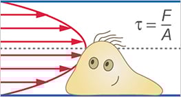
Shear stress is a force that acts on a surface when a solid object is pulled across the surface, or a liquid flows over it. It acts parallel to the surface, in the direction in which the object (or fluid) is moving. Shear stress (τ) is defined as force per area (τ = F/A, where τ = the shear stress, F = the force applied, and A = the cross-sectional area of the material surface).
Shear stress in fluids/flow profile: A fluid flowing through a channel can be seen as a stack of fluid layers moving on top of each other. The shear stress is caused by the friction between the layers, due to the fluid’s viscosity. The friction between the layers results in the velocity distribution, the flow profile. The viscosity determines how high the friction between the layers is.
Shear rate is defined as the change in velocity, at which one fluid moves over an adjacent layer. The shear rate is determined by both the vessel cross-section and the flow rate, measured in reciprocal seconds (s-1). The shear rate is an important parameter in rolling adhesion experiments, as it indicates how fast the cells roll over the surface; or how long the contact time is to the adherent cells.
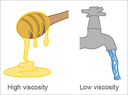
Viscosity (strictly speaking, dynamic viscosity) is the property of liquids that describes their fluidity. It decisively determines how easily liquid layers can slide over each other and is an important parameter for calculating shear stress. Water, for example, has a lower viscosity than honey. In Newtonian fluids (e.g., water, cell culture medium), the viscosity is independent of the flow rate.
Flow rate is the volume flow through a channel in a given time. The flow rate alone does not indicate the force applied to the adherent cells, but it is needed for the shear stress calculation. The flow rate (Φ) is defined as a volume per time (e.g., ml/min).
The Reynolds number (Re) describes whether a fluid flow is laminar or turbulent. The Reynolds number is given by the ratio of inertial forces to friction forces in a fluid. This value is dimensionless. Laminar flow occurs at low Reynolds numbers whereas turbulent flow occurs at high Reynolds numbers. The critical Reynolds number, which indicates laminar flow in pipes and biological vessels, is Re=2000. A Reynolds number of above approximately Re 4000 is most likely to represent a turbulent flow.
Flow Types Used to Apply Shear Stress
The flow type determines the wall shear stress pattern applied to adherent cells. Basically, flow types can be subdivided into laminar flow and turbulent flow. For most endothelial shear stress assays, unidirectional laminar flow is the flow type of choice, because it provides steady and reproducible wall shear stress. Pulsatile flow is useful when arterial-like dynamic flow conditions are required. Oscillatory flow is commonly used to model disturbed-flow environments, while non-uniform laminar flow is suitable for studying spatial shear stress differences, for example at vessel branching sites.
| Flow Type | Experimental Use | Biological Relevance | Flow Rate | Flow Direction | ibidi Compatibility |
|---|---|---|---|---|---|
| Unidirectional laminar flow | Endothelial cell investigation, barrier function, mechanotransduction, rolling and adhesion | Common, in many healthy vessels | Constant | Constant | ibidi Pump System, µ-Slide I Luer, µ-Slide VI |
| Non-uniform unidirectional laminar flow | Vessel branching models and spatial shear stress gradient studies | Occurs at vessel branching sites | Spatially variable across the cell layer | Constant | ibidi Pump System, µ-Slide y-shaped |
| Pulsatile laminar flow | Arterial-like flow models and dynamic vascular stimulation | Occurs in large arterial vessels due to heartbeat-driven fluctuations | Periodically changing | Constant | ibidi Pump System, µ-Slide I Luer, µ-Slide VI |
| Oscillatory laminar flow | Disturbed-flow models, endothelial dysfunction, inflammation-related assays | Used experimentally to model turbulent flow conditions | Constant | Periodically changing | ibidi Pump System, µ-Slide I Luer, µ-Slide VI |
| Turbulent flow | Conceptual reference; generally not used for controlled wall shear stress assays | Rare, associated with pathophysiological processes | Changing | Changing | No. Turbulent flow cannot be achieved in ibidi flow chambers using physiological flow regimes. Oscillatory laminar flow can be used to model turbulent flow conditions. |
Laminar Flow
Laminar flow is defined as the movement of liquids without turbulences. The fluid flows in parallel layers with no disruption between them. Laminar flow can be subdivided into the following:
- Unidirectional laminar flow (including non-uniform unidirectional laminar flow)
- Pulsatile laminar flow
- Oscillatory laminar flow
Unidirectional laminar flow is encountered in most small healthy biological vessels, such as small arteries and veins. In vivo, certain cells, such as endothelial cells and kidney epithelial cells, are constantly exposed to flow. Experimentally, unidirectional laminar flow is achieved by perfusing medium through low-walled channels, and by keeping both the flow direction and velocity constant over time.
Non-uniform unidirectional laminar flow: In this case, the flow direction is constant whereas the flow rate spatially varies across the cell layer. In vivo, a non-uniform laminar flow occurs at vessel branching sites.
Experimentally, non-uniform laminar shear stress can be achieved by a special channel geometry, which generates flow rate variations at specific sites within a slide. Using this experimental setup, different shear stresses on cells can be investigated using a single sample. This is an efficient way to investigate the effect of different shear stresses on cell physiology.
Pulsatile laminar flow is encountered in large arterial vessels due to the fluctuations caused by the heartbeat. Experimentally, this type of flow can be mimicked by employing a flow with a periodically changing flow rate while keeping the flow direction constant.
Oscillatory laminar flow is accepted as a means of simulating turbulences when using flow chambers. Although the flow is laminar, there is no main direction because the flow direction is changed at regular intervals (e.g., every 0.5 seconds). Besides during valve switching, the flow rate is kept constant.
A. Sabine, et al. FOXC2 and fluid shear stress stabilize postnatal lymphatic vasculature, The Journal of Clinical Investigation, 2015, 10.1172/JCI80454
read abstract

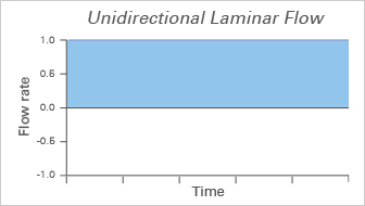
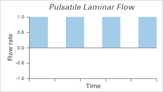
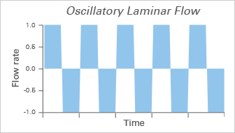
Laminar flow profiles. Arrows represent the distribution of velocities.
Turbulent Flow
Turbulent flow is characterized by unpredictable changes in both flow rate and direction over time. In vivo, turbulences are rare and can only be found during pathophysiological processes.
T. Hosoya, et al. Differential Responses of the Nrf2-Keap1 System to Laminar and Oscillatory Shear Stresses in Endothelial Cells. J Biol Chem, 2005, 10.1074/jbc.M502551200
read abstract
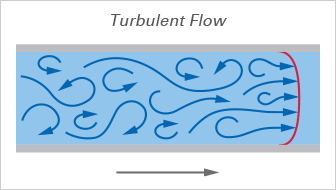
Turbulent flow profile. Arrows represent the distribution of velocities.
Principle
In vivo, endothelial cells develop and differentiate under shear stress conditions. When starting cell-based assays with endothelial cells, you should consider the possible influence of this mechanical force on cell morphology and physiology. To bring the cells to a more physiological, in vivo-like state, they are cultured under flow for hours up to several weeks, generating more relevant results. In other words, flow conditioning is important for studies using cells that are physiologically exposed to wall shear stress.
Application Examples
- Investigating the influence of shear stress on endothelial cell physiology with various experimental readouts, such as immunofluorescence, western blot, qPCR, and FACS
- Preparing the cell layer for subsequent functional assays, such as rolling and adhesion or transmigration assays
| Flow characteristics | Continuous laminar flow, Non-uniform flow, Oscillatory flow (needed for disturbed flow simulation), Pulsatile flow |
| Experiment duration | Hours, up to several weeks |
| Recommended pumps | ibidi Pump System |
| Recommended µ-Slides | µ-Slide I Luer Family, µ-Slide VI 0.4, µ-Slide y-shaped |


Human umbilical vein endothelial cells (HUVEC) cultured under flow conditions (20 dyn/cm²) in a µ-Slide I 0.4 Luer over 9 days. The primary cells were transduced with an adenoviral LifeAct vector 24 hours prior to the experiment.
Principle
In a rolling and adhesion assay, leukocytes and/or platelets are perfused over a surface with a protein coating or a cell layer. Their adhesion to and their interaction with the surface can be analyzed under various conditions (e.g., after gene knockdown or drug treatment).
Application Examples
- Investigating the adhesion of platelets and leukocytes on endothelial cells or matrix protein layers
Schulz C, et al. (2009) Novel Methods for Assessment of Platelet and Leukocyte Function Under Flow - Application of Epifluorescence and Two-Photon Microscopy in a Small Volume Flow Chamber Model. Open Biol J 2(1):130–136.
read abstract
| Flow characteristics | Continuous laminar flow |
| Experiment duration | Minutes to hours |
| Recommended pumps | ibidi Pump System, syringe pump, peristaltic pump |
| Recommended µ-Slides | µ-Slide I Luer Family, µ-Slide VI 0.4, µ-Slide y-shaped |
Endothelial Mechanotransduction
Wall shear stress is an important mechanical stimulus for endothelial cells. It can influence cytoskeletal organization, cell alignment, ion channel activation, gene expression, inflammatory signaling, and barrier function. Flow-based assays are therefore used to study how endothelial cells sense and respond to mechanical forces under more physiological conditions.
Disturbed Flow and Atherosclerosis Models
Disturbed or oscillatory flow conditions are used to model flow environments that occur at vessel branches or under pathophysiological conditions. These assays can help investigate inflammatory signaling, endothelial dysfunction, and shear stress-dependent mechanisms relevant to vascular disease models.
Plan Your ibidi Setup for Wall Shear Stress Assays
Wall shear stress assays require a defined flow source, a channel geometry with known dimensions, and stable environmental conditions during imaging or endpoint analysis. For a complete overview of compatible ibidi components and workflow considerations, see the detailed setup guide.
Frequently Asked Questions About Cells Under Shear Stress
What is wall shear stress?
Wall shear stress is the force generated at the boundary between a flowing liquid and a surface. In cell culture channels, it describes the mechanical force experienced by adherent cells at the channel surface and can influence cell morphology, adhesion, signaling, gene expression, and physiological behavior.
Why is shear stress important in cell culture?
Shear stress is an important physical stimulus in many biological systems. In standard static in vitro culture, this mechanical stimulus is missing. Applying defined shear stress can help create more physiologically relevant conditions for cells that naturally experience flow in vivo.
Which cell types are commonly studied under shear stress?
Common examples include vascular endothelial cells, lymphatic endothelial cells, epithelial cells from organs such as the kidney or lung, and cells involved in rolling and adhesion assays, such as leukocytes or platelets. The relevance depends on whether the cell type naturally experiences fluid flow or interacts with surfaces under flow conditions.
What is the difference between laminar and turbulent flow?
Laminar flow is characterized by ordered liquid movement in parallel layers, while turbulent flow shows unpredictable changes in flow rate and direction. In many cell culture flow assays, laminar flow is used because it allows better control of the shear stress applied to the cells.
What is the difference between unidirectional, pulsatile, and oscillatory flow?
Unidirectional flow has a constant direction and can provide steady wall shear stress. Pulsatile flow has a periodically changing flow rate while the direction remains constant, resembling dynamic arterial conditions. Oscillatory flow changes direction periodically and is often used to model disturbed-flow conditions.