


Applications of Wall Shear Stress Assays
This page summarizes common wall shear stress assay applications and cell culture under flow applications for adherent cells exposed to defined mechanical stimulation under controlled in vitro conditions. It is intended for researchers planning endothelial, epithelial, vascular co-culture, immune cell interaction, and mechanotransduction assays under flow.
Depending on the biological question, cells can be cultured as a monolayer on a coverslip, on an extracellular matrix or gel matrix, in or adjacent to a gel matrix, or as co-culture monolayers separated by an optical porous membrane. This page connects these assay formats with experimental examples, typical readouts, and suitable ibidi solutions.
In brief: Wall shear stress assays are used to study how adherent cells respond to defined fluid flow. Major applications include endothelial cell conditioning, mechanotransduction, disturbed-flow models, extracellular matrix-based flow assays, cells in or on gel matrices, co-culture models on porous membranes, rolling and adhesion assays, immunofluorescence analysis, and impedance measurements under flow.
Learn the biological background of wall shear stress and flow types, or plan the technical setup with the ibidi setup guide for wall shear stress assays.

Application Overview
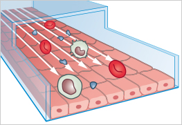
Different wall shear stress assay formats can be used to simulate mechanical forces generated by fluid flow in biofluidic systems, such as blood vessels, lymphatic vessels, and epithelial channels. The choice of assay format depends on the cell type, substrate, extracellular matrix, co-culture requirements, flow profile, experiment duration, and readout method.

| Application | Assay Principle | Best Used For | ibidi Solutions |
|---|---|---|---|
| Cell Monolayer on Coverslip | Adherent cells are cultured as a monolayer on an optically suitable surface and exposed to defined wall shear stress. | Endothelial cell conditioning, mechanotransduction, disturbed-flow models, immunofluorescence, impedance measurements, and rolling and adhesion assays | ibidi Pump System, µ-Slide I Luer Family, µ-Slide VI, µ-Slide y-shaped |
| Cell Monolayer on Gel Matrix | Cells are cultured under flow on a defined extracellular matrix or gel matrix. | Matrix-dependent adhesion, extracellular matrix signaling, endothelial alignment, cytoskeletal organization, and matrix-dependent responses to shear stress | ibidi Pump System, µ-Slide I Luer 3D, Collagen Type I |
| Cell Monolayer on Gel Matrix: Cells in Flow / Inside a Gel Matrix | Cells are cultured on a gel matrix or embedded inside a gel matrix while flow is applied through an adjacent or connected channel. | Matrix-rich or 3D-like microenvironments, cell–matrix interactions, matrix remodeling, and gel-based endothelial or epithelial flow models | ibidi Pump System, µ-Slide I Luer 3D, Collagen Type I |
| Co-Culture of Cell Monolayers on an Optical Porous Membrane | Two cell layers are cultured in separate but interacting compartments, with one side exposed to flow. | Barrier models, endothelial–epithelial interaction studies, immune cell interaction studies, compartmentalized culture, and microscopy-based co-culture analysis under flow | ibidi Pump System, µ-Slide ibiPore SiN |
Which Wall Shear Stress Assay Format Should You Choose?
The optimal assay format depends on whether the experiment focuses on direct shear stress exposure, matrix-dependent cell behavior, a gel-based microenvironment, or compartmentalized co-culture. The table below provides a practical starting point for selecting the most suitable format.
| Experimental Need | Recommended Format | Reason |
|---|---|---|
| Standard endothelial monolayer under defined shear stress | Cell Monolayer on Coverslip | Provides direct flow exposure, optical access, and compatibility with long-term flow conditioning, microscopy, staining, impedance measurements |
| Matrix-dependent adhesion or collagen-based culture | Cell Monolayer on Gel Matrix | Combines wall shear stress with extracellular matrix-dependent cell attachment and signaling |
| Matrix-rich or 3D-like microenvironment | Cell Monolayer under Flow: Cells in Flow / Inside a Gel Matrix | Allows flow exposure in combination with gel-based cell culture, matrix remodeling, and cell–matrix interactions, rolling and adhesion assays |
| Barrier models or interacting cell layers | Co-Culture of Cell Monolayers on an Optical Porous Membrane | Enables compartmentalized culture with flow exposure and microscopy-compatible membrane-based analysis |
Cell Monolayer on Coverslip
In this assay format, adherent cells are cultured as a monolayer on an optically suitable surface and exposed to defined wall shear stress. This format is commonly used for endothelial cell conditioning, live cell imaging, immunofluorescence staining, impedance-based monitoring, rolling and adhesion assays, mechanotransduction studies, and disturbed-flow models.
| Best suited for | Direct shear stress exposure of endothelial or epithelial monolayers, long-term flow conditioning, microscopy-based analysis, impedance measurements, and rolling or adhesion assays |
| Typical readouts | Cell morphology, alignment, immunofluorescence, live cell imaging, impedance, qPCR, western blot, FACS, rolling velocity, and adhesion frequency |
| Recommended setup | ibidi Pump System, µ-Slide I Luer Family, µ-Slide VI, µ-Slide y-shaped |
Endothelial Cell Conditioning Under Shear Stress
In vivo, vascular endothelial cells are continuously exposed to wall shear stress generated by blood flow. In static cell culture, this mechanical stimulus is missing. Long-term flow conditioning of endothelial cell monolayers helps induce a more physiological phenotype and can influence cell morphology, alignment, cytoskeletal organization, adhesion properties, gene expression, and barrier function. This assay format is commonly used to investigate endothelial cell physiology, mechanotransduction, inflammatory activation, and the preparation of cell layers for subsequent functional assays such as rolling and adhesion or transmigration assays.
| Flow characteristics | Unidirectional laminar flow, pulsatile flow, oscillatory flow, non-uniform flow |
| Experiment duration | Hours to several weeks |
| Typical readouts | Cell morphology, alignment, immunofluorescence, western blot, qPCR, FACS, barrier-related markers |
| Recommended pump | ibidi Pump System |
| Recommended µ-Slides | µ-Slide I Luer Family, µ-Slide VI, µ-Slide y-shaped |
Phase Contrast and Fluorescence Microscopy of Pulmonary Endothelial Cells (HPMECs)
When cultured under flow, human pulmonary microvascular endothelial cells (HPMECs) show changes in morphology, cytoskeletal organization, and endothelial cell–cell contacts compared with static culture. This example demonstrates how phase contrast and fluorescence microscopy can be used to assess endothelial responses to defined shear stress conditions.
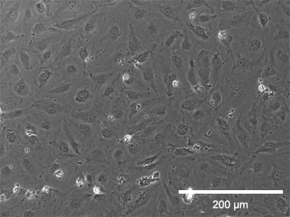
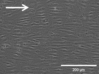

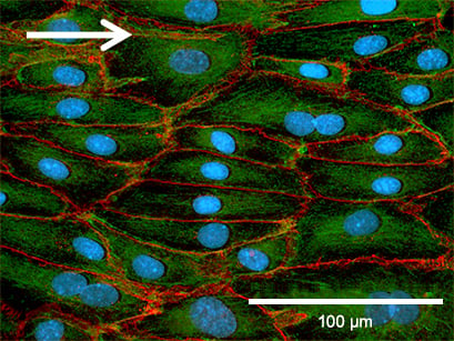
Phase contrast microscopy (top, scale bar: 200 µm) and fluorescence microscopy (bottom, scale bar: 100 µm) of HPMECs comparing static culture (left) and flow culture (right) after 72 h. Fluorescence labels: β-actin (green), VE-cadherin (red), and DAPI (blue). Data by Daniel Bourquain, Robert Koch Institute, Berlin.
Immunofluorescence of Flow-Conditioned Endothelial Cells
Immunofluorescence staining allows the comparison of static and flow-cultured endothelial cells and can reveal differences in cytoskeletal organization, adherens junctions, tight junctions, Golgi orientation, endothelial markers, and overall monolayer organization. In these examples, flow-conditioned HUVECs are compared with static cultures using different markers. Together, these stainings illustrate how wall shear stress affects endothelial cell shape, junctional organization, polarity, and marker distribution.
VE-cadherin staining highlights adherens junctions in both static and flow-conditioned HUVECs. Under static conditions, the cells are generally larger and show a less organized actin cytoskeleton. In contrast, flow-conditioned cells are elongated and display distinct F-actin stress fibers, indicating flow-dependent reorganization of the endothelial cell layer.

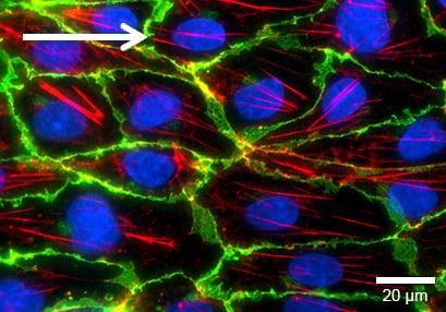
HUVECs were cultured for 5 days under static conditions in a µ-Dish 35 mm ibiTreat (left, 0 dyn/cm²) or under flow at 10 dyn/cm² (right) in a µ-Slide I 0.4 Luer ibiTreat. The cells were stained for VE-cadherin (green), F-actin (red), and nuclei (blue). Imaging was performed using a Nikon Eclipse microscope at 60× magnification.
Claudin-5, a tight junction protein located at endothelial cell–cell contacts, becomes clearly visible at contact zones in flow-conditioned HUVECs after several days of shear stress exposure. This staining shows how flow conditioning can support endothelial differentiation and junctional organization.
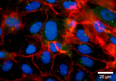
HUVECs were cultured for 5 days under static conditions in a µ-Dish 35 mm ibiTreat (left, 0 dyn/cm²) or under flow at 10 dyn/cm² (right) in a µ-Slide I 0.4 Luer ibiTreat. The cells were stained for claudin-5 (green), F-actin (red), and nuclei (blue). Imaging was performed using a Nikon Eclipse microscope at 60× magnification.
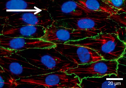
Human Golgin-97 staining can be used to visualize the Golgi apparatus as a marker for cell polarity and intracellular organization. In flow-conditioned HUVECs, the Golgi apparatus is localized along the direction of flow, supporting the analysis of flow-induced polarization within endothelial cells.
von Willebrand factor (vWF) is a characteristic endothelial marker. Under flow, vWF multimers can elongate into rod-like structures associated with the cell membrane, illustrating how shear stress can influence endothelial marker organization and flow-dependent endothelial cell physiology.
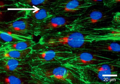
HUVECs were cultured under flow at 10 dyn/cm² for 4 days in a µ-Slide I 0.4 Luer ibiTreat. Cells were stained for human Golgin-97 (red), F-actin (green), and nuclei (blue). Imaging was performed using a Nikon Eclipse microscope at 60× magnification.

HUVECs were cultured under flow at 10 dyn/cm² for 5 days in a µ-Slide I 0.4 Luer ibiTreat. The cells were stained for von Willebrand factor (green), and nuclei were stained with DAPI (blue). Imaging was performed using a Nikon Eclipse microscope at 60× magnification.
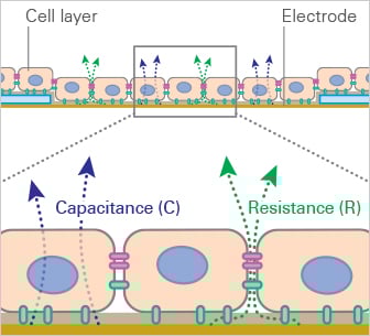
Impedance Measurements Under Flow Stimulation
Mechanical perturbations have a profound effect on the characteristics of cell layers in vitro. In long-term experiments with HUVEC, three different phenotypes can be observed: a round flat cell after seeding, elongated cells after 1–2 days, and finally an oriented cobblestone appearance of a dense, compact cell layer. The morphological changes are accompanied by physiological changes of the endothelial cell monolayer, which can be measured by impedance monitoring.
Cells are cultivated in channels with electrode arrays. Depending on the state of development of the endothelial cell monolayer, the gaps between the cells (influenced by the cell–cell contacts) change in size. These shifting gaps can be characterized by their change in conductivity when applying AC currents of different frequencies. The continuous red lines in the scheme represent the ion currents between the cells when applying the AC current.
Static culture

After seeding, cells grow to confluence within 2–3 days. During this time, conductivity decreases and resistance rises to a plateau, which is then maintained under subsequent static culture conditions for several days.
Flow culture
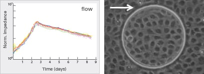
Applying shear stress reduces conductivity and increases the impedance of the monolayer. Over several days, the impedance of the endothelial monolayer decreases, indicating altered physiological properties compared with static culture conditions.
Disturbed Flow and Atherosclerosis Models
Disturbed-flow and oscillatory-flow assays are used to model flow environments that occur at vessel branches or under pathophysiological vascular conditions. In contrast to steady unidirectional laminar flow, oscillatory flow changes direction over time and can be used to investigate endothelial dysfunction, inflammatory signaling, oxidative stress responses, and shear stress-dependent mechanisms relevant to vascular disease models such as atherosclerosis.
Hosoya T, et al. (2005) Differential responses of the Nrf2-Keap1 system to laminar and oscillatory shear stresses in endothelial cells. J Biol Chem 280(29):27244–27250. 10.1074/jbc.M502551200.
Read article
| Flow characteristics | Oscillatory laminar flow, non-uniform laminar flow |
| Experiment duration | Hours to several days |
| Typical readouts | Inflammatory signaling, oxidative stress response, endothelial dysfunction markers, gene expression |
| Recommended pump | ibidi Pump System |
| Recommended µ-Slides | µ-Slide I Luer Family, µ-Slide VI, µ-Slide y-shaped |
Many endothelial and epithelial cells interact with extracellular matrix proteins in vivo. Culturing a cell monolayer under flow on a defined matrix, such as Collagen I, makes it possible to combine wall shear stress with matrix-dependent cell adhesion and signaling. This assay format is useful for analyzing cell morphology, cytoskeletal organization, adhesion, endothelial alignment, and matrix-dependent responses under physiologically relevant flow conditions. It can be combined with phase contrast microscopy, live cell imaging, immunofluorescence staining, and endpoint analysis after flow conditioning.
| Best suited for | Matrix-dependent adhesion, extracellular matrix signaling, endothelial alignment, cytoskeletal organization, and matrix-dependent responses to wall shear stress |
| Typical readouts | Phase contrast microscopy, live cell imaging, immunofluorescence, cell morphology, cytoskeletal organization, and endpoint analysis |
| Recommended setup | ibidi Pump System, µ-Slide I Luer 3D, Collagen Type I |
Cell Monolayer on Gel Matrix: Cells in Flow / Inside a Gel Matrix
Flow assays can also be performed with cells cultured on a gel matrix or with cells embedded inside a gel matrix. This approach combines wall shear stress, extracellular matrix interaction, and three-dimensional cell–matrix organization. It is especially useful for studying endothelial or epithelial responses in more complex microenvironments, including matrix remodeling, cell morphology, cell behavior, and signaling under flow. Depending on the assay design, cells can be exposed directly to flow at the gel surface or cultured within a matrix while flow is applied through an adjacent channel.
| Best suited for | Rolling and adhesion assays, matrix-rich or 3D-like microenvironments, cell–matrix interaction studies, matrix remodeling, and gel-based endothelial or epithelial flow models |
| Typical readouts | Cell morphology, cell behavior, matrix interaction, immunofluorescence, live cell imaging, and endpoint staining |
| Recommended setup | ibidi Pump System, µ-Slide I Luer 3D, Collagen Type I |
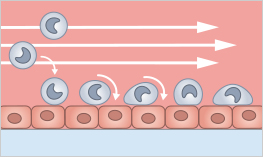
Rolling and Adhesion Assays
Rolling and adhesion assays are used to investigate how leukocytes, platelets, or other suspended cells interact with a protein-coated surface or an adherent cell layer under defined flow conditions. In this assay, cells are perfused through a channel while their rolling behavior, adhesion frequency, interaction time, and response to experimental treatments such as gene knockdown, inflammatory stimulation, or drug exposure are analyzed by microscopy. The assay is especially relevant for vascular inflammation, immune cell recruitment, platelet adhesion, and endothelial interaction studies.
Schulz C, et al. (2009) Novel methods for assessment of platelet and leukocyte function under flow: application of epifluorescence and two-photon microscopy in a small volume flow chamber model. Open Biol J 2(1):130–136. 10.2174/1874196700902010130.
Read article
| Flow characteristics | Unidirectional laminar flow |
| Experiment duration | Minutes to hours |
| Typical readouts | Rolling velocity, adhesion frequency, cell–surface interaction, platelet or leukocyte behavior |
| Recommended pumps | ibidi Pump System, syringe pump, peristaltic pump |
| Recommended µ-Slides | µ-Slide I Luer Family, µ-Slide VI, µ-Slide y-shaped |
Co-culture flow assays on an optical porous membrane enable the cultivation of two cell layers in separate but interacting compartments. One cell monolayer can be exposed to flow while soluble factors, cell–cell communication, or barrier-related responses are analyzed across the membrane. This assay format is suitable for endothelial and epithelial barrier models, vascular co-culture systems, immune cell interaction studies, and live or fixed-cell microscopy of cell layers under defined shear stress conditions.
| Best suited for | Barrier models, endothelial–epithelial interaction studies, immune cell interaction studies, compartmentalized culture, and microscopy-based co-culture analysis under flow |
| Typical readouts | Barrier function, cell–cell interaction, immunofluorescence, phase contrast microscopy, and live cell imaging |
| Recommended setup | ibidi Pump System, µ-Slide ibiPore SiN |
Plan Your ibidi Setup for Wall Shear Stress Assays
Wall shear stress assays require a defined flow source, a channel geometry with known dimensions, and stable environmental conditions during imaging or endpoint analysis. For a complete overview of compatible ibidi components and workflow considerations, see the detailed setup guide.
Frequently Asked Questions About Wall Shear Stress Assay Applications
Which wall shear stress assay format is best for endothelial monolayers?
For standard endothelial monolayers under defined shear stress, a cell monolayer on a coverslip or optical bottom is usually the most direct assay format. It allows controlled flow exposure, microscopy access, long-term flow conditioning, immunofluorescence staining, impedance measurements, and downstream endpoint analysis.
When should I use a gel matrix in a wall shear stress assay?
A gel matrix is useful when matrix-dependent adhesion, extracellular matrix signaling, or a more tissue-like microenvironment is relevant to the biological question. It can be used for cell monolayers on a matrix or for cells cultured in or adjacent to a gel matrix.
When should I use a cells-in-flow or inside-gel-matrix format?
This format is suitable when the experiment should combine defined flow with a matrix-rich or 3D-like microenvironment. It can support studies of cell–matrix interactions, matrix remodeling, cell morphology, and endothelial or epithelial responses in gel-based culture models.
When should I use a porous membrane co-culture format?
A porous membrane co-culture format is useful when two cell layers should be cultured in separate but interacting compartments. It is suitable for barrier models, endothelial–epithelial interaction studies, immune cell interaction studies, and microscopy-based analysis under flow.
Which ibidi products are commonly used for wall shear stress assay applications?
Common products include the ibidi Pump System, µ-Slide I Luer Family, µ-Slide VI, µ-Slide y-shaped, µ-Slide I Luer 3D, µ-Slide ibiPore SiN, and compatible matrices such as Collagen Type I. The optimal setup depends on the assay format, flow profile, cell type, matrix requirements, co-culture design, and readout method.