



Outstanding Research
in Neurobiology
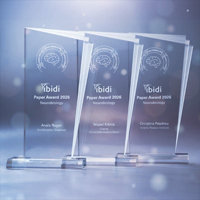
We are pleased to announce the winners of the ibidi Paper Award 2026, honoring outstanding contributions to neurobiology research. Each awardee will receive €500 in recognition of their excellent scientific work.
Congratulations to the 2026 Award Winners!
We sincerely thank everyone who submitted their work and contributed to this year’s award. Our special thanks go to our jury members, Henrietta M. Nielsen, PhD, Moran Amit, M.D., Ph.D., and Rosa Chiara Paolicelli, PhD, for their time, expertise, and thoughtful evaluation.
Anaïs Roger for Her Publication in Immunity (IF: 26.3)
Sensory neuron production of substance P and TAFA4 promotes disease tolerance during viral infection
Anaïs Roger earned both her MSc and PhD in Immunology from Aix-Marseille University, where she conducted her doctoral research at the Centre d’Immunologie de Marseille-Luminy, France, under the supervision of Dr. Sophie Ugolini and was supported by the Fondation pour la Recherche Médicale.
Her doctoral work focused on the role of sensory neurons in regulating immunity during cutaneous HSV-1 infection. By combining advanced in vitro and in vivo approaches, she demonstrated that virus-activated sensory neurons promote disease tolerance through the release of the neuropeptides Substance P and TAFA4. This release limits harmful inflammation and supports tissue protection.
Currently, Anaïs Roger is a postdoctoral fellow at Queen’s University, Canada, under the supervision of Dr. Sébastien Talbot. Her current research explores neuroimmune interactions in the bone marrow and how the nervous system shapes immune responses in health and disease.
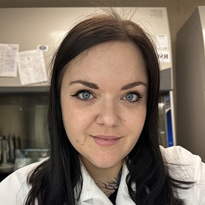
Anaïs Roger, Aix-Marseille University, France / Queen’s University, Canada
Why Anaïs’s Publication Convinced the Jury—Moran Amit
Roger et al. stands out for its elegant resolution of a fundamental question in neuroimmune biology: how sensory neurons actively choreograph immune cell migration during viral infection. Using rigorous in vitro chemotaxis assays and in vivo mixed bone marrow chimera models, the authors demonstrate that Substance P, released by TRPV1+ nociceptors, directly inhibits CXCL1-driven neutrophil migration through the MRGPRA1 receptor, redefining Substance P as an anti-inflammatory, pro-tolerance signal.
The mechanistic clarity, multi-layered genetic and pharmacological validation, and translational implications for infection-associated immunopathology make this an outstanding and timely contribution to the field of cell migration research.
Publication
Roger, A., et al. (2026). Sensory neuron production of substance P and TAFA4 promotes disease tolerance during viral infection. Immunity, 59(3), 682-699.e8.
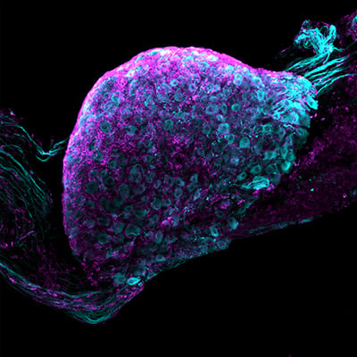
Whole-mount dorsal root ganglion (DRG) preparation imaged in a µ-Dish 35 mm, high, showing βIII-tubulin–positive neurons (cyan) and immune cells (magenta). The image highlights robust immune cell infiltration within the DRG following HSV-1 infection, illustrating neuroimmune interactions at the level of sensory ganglia.
Majed Kikhia for His Publication in Nature Communications (IF: 15.7)
Multicolor fate mapping of microglia reveals polyclonal proliferation, heterogeneity, and cell-cell interactions after ischemic stroke in mice
Majed studied medicine in Syria and earned a Master’s degree in Medical Neurosciences from the Charité – Universitätsmedizin Berlin, where he is currently an M.D./Ph.D. candidate and a fellow of the Einstein Center for Neurosciences Berlin.
In his doctoral work, he explored the responses of microglia and other myeloid cells to ischemic stroke using multicolor fate mapping models. Majed is a passionate microscopist at heart who enjoys witnessing natural phenomena at the cellular and subcellular levels. For example, he applied various microscopy techniques to demonstrate microglial cell-to-cell interactions in ischemic brain tissue.
In his work, he aims to combine rigorous experimental approaches with quantitative computational methods to advance the knowledge of microglial heterogeneity, morphology, and self-organization, as well as glial-neuronal and glial-glial interactions in health and disease.
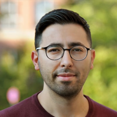
Majed Kikhia, Charité – Universitätsmedizin Berlin, Germany
Why Majed’s Publication Convinced the Jury—Rosa Chiara Paolicelli
In this study, Kikhia et al. explore microglial heterogeneity after ischemic stroke using multicolor fate mapping in combination with ex vivo and in vivo imaging. The authors elegantly show that stroke induces microglia polyclonal proliferation with a distinct temporal course. They further show that clonal identity contributes to microglial heterogeneity within the ischemic tissue, with cells of the same clone exhibiting similar electrophysiological and morphological profiles.
One of the most exciting findings of the study is the close interaction between microglia from different clones, which would have been impossible to appreciate without the multicolor fate mapping approach. This interesting study advances the current understanding of microglial heterogeneity in response to stroke and helps visualize how these cells dynamically interact with one another within the ischemic lesion.
Publication
Kikhia, M., et al. (2025). Multicolor fate mapping of microglia reveals polyclonal proliferation, heterogeneity, and cell-cell interactions after ischemic stroke in mice. Nature Communications, 16(1), 8294.
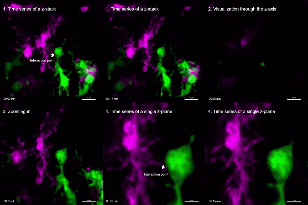
Live-cell imaging of microglial process-soma interaction in the ischemic tissue of an acute brain slice 8 weeks after stroke, imaged in a µ-Dish 35 mm, high. The RFP+ cell (magenta) extends a process (white arrowhead) to establish contact with the soma of a YFP+ cell (green) and retracts it later
Christina Paschou for Her Publication in PNAS (IF: 9.1)
Dysregulated proteostasis in p.A53T-α-Synuclein astrocytes aggravates Lewy-like neuropathology in a Parkinson's disease iPSC model
Christina is a Ph.D. candidate in Neurobiology at the Hellenic Pasteur Institute, Athens, Greece, where she studies astrocyte involvement in neurodegenerative diseases using human induced pluripotent stem cell (iPSC)-based models. She holds a B.Sc. in Biological Applications and Technologies from the University of Ioannina, where her undergraduate research focused on astrocytes in the substantia nigra of neonatal postmortem brain tissue, which sparked her interest in neuroscience.
She later completed the Athens International Master’s Program in Neurosciences, during which she was trained in pluripotent stem cell-derived models of human astrocytes. Her doctoral research focuses on developing and applying iPSC-derived midbrain astrocyte-dopaminergic neuron co-culture systems carrying the Parkinson’s disease-linked A53T α-synuclein mutation.
Her work has demonstrated that astrocyte dysfunction contributes to neuronal vulnerability and disease pathogenesis. Christina has expertise in stem cell technologies, electron and confocal microscopy, and image analysis. Her research aims to elucidate astrocyte-mediated mechanisms in neurodegeneration.
Christina Paschou, Hellenic Pasteur Institute, Athens, Greece
Why Christina’s Publication Convinced the Jury—Henrietta M. Nielsen
Parkinson’s disease is characterized by the loss of dopaminergic neurons in the substantia nigra and the accumulation of alpha-synuclein in Lewy bodies. However, the direct contribution of astrocytes to Parkinson’s disease pathology remains insufficiently understood. Using patient-derived iPSCs and induced astrocytes carrying the p.A53T SNCA mutation, together with mutation-corrected and healthy controls, Paschou and colleagues show that astrocytes express SNCA and produce measurable alpha-synuclein, including phosphorylated p-Ser129 alpha-synuclein.
Levels were increased in mutation-carrying astrocytes, which also displayed intrinsic defects in calcium homeostasis, lysosomal function, and alpha-synuclein clearance. In an elegant astrocyte-neuron co-culture system, mutant astrocytes induced Parkinson’s disease-associated pathology in healthy neurons, while healthy astrocytes showed protective effects on mutant neurons. By using human astrocytes, the study provides highly translation-relevant insights into astrocyte-driven mechanisms in Parkinson’s disease and highlights astrocytes as potential therapeutic targets in Parkinson’s disease pathogenesis and progression.
Publication
Paschou, C., et al. (2025). Dysregulated proteostasis in p.A53T-α-Synuclein astrocytes aggravates Lewy-like neuropathology in a Parkinson's disease iPSC model. Proceedings of the National Academy of Sciences of the United States of America, 122(47), e2505240122.
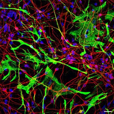
Confocal image of a 14-day co-culture of Parkinson’s disease (PD) iPSC-derived astrocytes and neurons. Astrocytes were immunostained for GFAP (green), and neurons for MAP2 (red). Scale bar, 20 μm.
Henrietta M. Nielsen, PhD
Stockholm University, Sweden
Dr. Henrietta M. Nielsen received her PhD in Medicine from Lund University, Sweden, and completed postdoctoral training at VU Medical Center, Amsterdam, the Netherlands, and at Lund University. Before starting her independent career as Assistant Professor of Neurochemistry at Stockholm University, she also trained at Mayo Clinic Florida, USA.
Dr. Nielsen is a tenured Associate Professor in Neurochemistry. Her laboratory investigates biological mechanisms that promote and contribute to neurodegenerative dementia, with a major focus on peripheral biology. In particular, the Nielsen lab aims to understand how the APOE4 allele increases the risk of neurodegenerative diseases such as Alzheimer’s disease, with a strong interest in defining APOE4-associated peripheral phenotypes, especially in the liver. Dr. Nielsen serves as Senior Editor for Molecular Neurodegeneration and is an Editorial Board Member of Aging Cell. She is a Board Member of the International Society for Molecular Neurodegeneration (ISMND) and currently serves as ISMND President. Since 2024, Dr. Nielsen has been part of the Executive Organizing Team of the ADPD conference series.
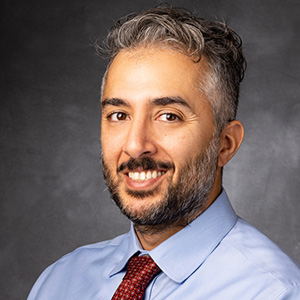
Moran Amit, M.D., Ph.D.
The University of Texas MD Anderson Cancer Center, USA
Dr. Moran Amit is a physician-scientist and principal investigator at The University of Texas MD Anderson Cancer Center, where he leads research at the interface of neurobiology, cancer biology, and immunology. He is a pioneer of cancer neuroscience, a rapidly emerging field that investigates how the nervous system contributes to tumor initiation, progression, and therapy response. His work focuses on neuro-tumor interactions in solid cancers, particularly head and neck malignancies and peripheral nervous system involvement.
Dr. Amit’s laboratory develops experimental models to study neural niche subpopulations and neurobiological signaling pathways within the tumor microenvironment. His research has revealed how cancer cells actively remodel neural circuits and induce nerve injury, thereby influencing inflammation, immune exhaustion, and resistance to immunotherapy. These discoveries have provided new insights into neurobiological mechanisms underlying tumor invasion, metastasis, and treatment failure.
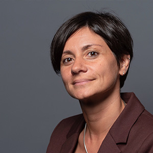
Rosa Chiara Paolicelli, PhD
University of Lausanne, Switzerland
Rosa obtained a Bachelor’s degree in Medical Biotechnology from the University of Bologna, Italy, followed by a Master’s in Molecular Neuroscience at the University of Bristol, UK. She then completed a PhD in Cellular and Molecular Biology at the European Molecular Biology Laboratory (EMBL), before undertaking postdoctoral training at the Institute of Regenerative Medicine at the University of Zurich.
Since 2018, Rosa has been a member of the Department of Biomedical Sciences at the University of Lausanne, where she is currently an associate professor and head of the Microglia Biology Lab. Her research explores the molecular and cellular mechanisms that regulate microglial function in health and disease, with a particular focus on genetic risk factors and metabolic control. Combining complementary in vitro and in vivo approaches, her lab investigates how microglial dysfunction contributes to brain development and neurodegeneration.
Check out the winners of the ibidi Paper Award 2025 (Cell Migration) here.