


Immunofluorescence Using ibidi Labware
Perform reliable immunofluorescence staining with optimized workflow guidance. This guide explains the differences between ibidi Chambered Coverslips, Channel Slides, and removable Chamber Slides. Learn how to select the right format based on microscopy setup, reagent volume, handling requirements, and sample storage. Explore labware comparison, benefits, and protocol differences.
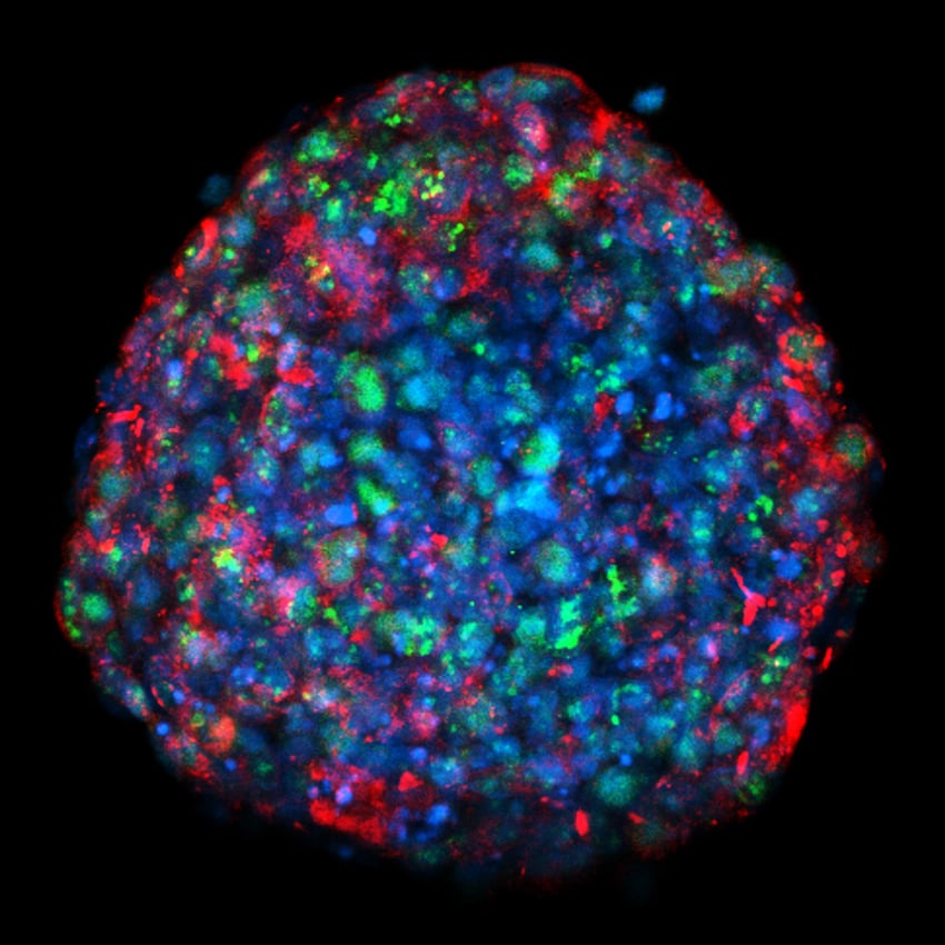
Confocal MIP image of an 8-day breast tumor spheroid mounted on an ibidi µ-Slide 18 Well Glass Bottom. The image shows stainings for nuclei (blue), Ki67+ cells (green) and gap junction protein (red). Image by Marina Rodriguez-Candela Mateos, Institute of Biomedical Research of A Coruña (INIBIC), A Coruna, Spain.
Benefits of ibidi µ-Chambers
- High-resolution imaging
Ideal for widefield fluorescence, confocal imaging, FRAP, FRET, FLIM, and undisturbed phase contrast imaging. - Fast and simple handling
All-in-one chambers simplify your immunofluorescence workflow. - Cost-effective experiments
Requires only a small number of cells and low reagent volumes.
ibidi Mounting Medium
High-quality non-hardening mounting medium with or without DAPI, optimized for fluorescence microscopy.
Comparison of Immunocytochemistry Protocols:
Traditional Staining vs. Staining With ibidi Labware
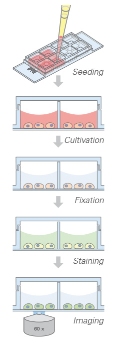
Using ibidi labware significantly shortens the immunofluorescence workflow compared to traditional coverslip methods. Cells can be cultured and stained directly in µ-Slides, eliminating additional handling steps and reducing protocol time. Explore the step-by-step workflow for traditional immunofluorescence staining using coverslips in comparison to the step-by-step workflow for staining directly in ibidi µ-Slides, minimizing handling and reducing overall assay time.
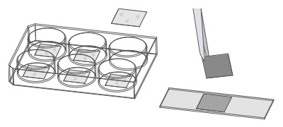
Protocol With Cells on Coverslips
Traditional method with nail polish mounting
| Step | Traditional Coverslip Workflow |
|---|---|
| 1 | Sterilize coverslips |
| 2 | Coat coverslips |
| 3 | Place sterile, coated coverslips into 6 well plate |
| 4 | Seed cells in large volume |
| 5 | Peel off the coverslip |
| 6 | Wash |
| 7 | Fix → Wash → Permeabilize → Wash → Block |
| 8 | Incubate in primary antibody → Wash → Incubate in secondary antibody → Wash |
| 9 | Mount cells with mounting medium |
| 10 | Mount coverslip with nail polish |
Protocol With ibidi µ-Slides
Time-saving method using all-in-one chambers
| Step | ibidi µ-Slide Workflow |
|---|---|
| 1 | Skip |
| 2 | Skip |
| 3 | Skip |
| 4 | Seed cells directly in chamber (low volume) |
| 5 | Skip |
| 6 | Wash |
| 7 | Fix → Wash → Permeabilize → Wash → Block |
| 8 | Incubate in primary antibody → Wash → Incubate in secondary antibody → Wash |
| 9 | Mount cells with mounting medium |
| 10 | Skip |
ibidi Labware for Immunofluorescence Compared
The table below provides a quick comparison of key immunofluorescence labware features.
| Feature | Chambered Coverslips | Channel Slides | Removable Chamber Slides | Traditional 6 Well Plate |
|---|---|---|---|---|
| Product example | µ-Slide 8 Well high | µ-Slide VI | 8 Well Chamber, removable | Standard 6 well plate (not offered by ibidi) |
| Bottom material | #1.5H Glass Coverslip or #1.5 Polymer Coverslip | #1.5H Glass Coverslip or #1.5 Polymer Coverslip | Standard glass slide | Polymer |
| IF staining workflow | Seed, stain, and image directly in chambered coverslip | Seed, stain, and image directly in channel slide | Seed and stain on glass slide, mount with coverslip for imaging | Seed and stain on coverslip, mount on glass slide for imaging |
| Number of IF protocol steps | Few | Few | Few | Many |
| High-throughput stainings | Limited | Limited | Yes | No |
| Low-volume stainings | Yes | Yes | Limited | No |
| Homogeneous cell & antibody distribution | Limited | Yes | Limited | No |
| Additional coverslips required | No | No | Yes | Yes |
| Typical mounting medium | Non-hardening | Non-hardening | Hardening | Non-hardening |
| Microscope type | Inverted | Inverted | Inverted & upright | Inverted & upright |
| Sample storage | Short-term | Short-term | Long-term | Long-term |
| Typical use | Parallel immunofluorescence assays without coverslip handling | Low-volume immunofluorescence staining with precise medium exchange | Mounted samples for long-term storage or staining jar workflows | Low-budget IF staining when time is not critical |
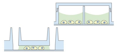
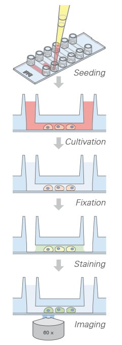
Chambered Coverslips
Chambered Coverslips allow performing the entire staining protocol without additional coverslips, with imaging directly through the coverslip bottom.
Advantages
- 1 to 18 non-removable wells on a coverslip bottom
- No coverslip handling
- Parallel assays without cross-contamination
Limitations
- Storage limited to weeks due to gas exchange through plastic
Channel Slides
Channel Slides are ideal for low-volume staining with precise medium exchange. The coverslip bottom eliminates the need for additional coverslips.
Advantages
- Different channel heights and coatings available
- No coverslip handling
- Low volumes of reagents
- Homogeneous cell and antibody distribution
Limitations
- Storage limited to weeks due to gas exchange through plastic
Removable Chamber Slides
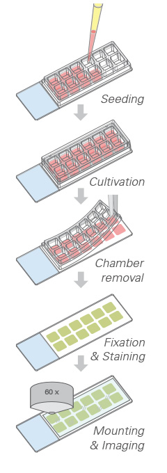
Removable Chamber Slides feature a silicone gasket mounted on a glass slide and are ideal for long-term storage of samples mounted with a coverslip.
Advantages
- Removable silicone chambers on a standard glass slide
- Ideal for long-term storage
- High-throughput screening possible
Limitations
- No high-resolution microscopy during cell cultivation
FAQ
Which ibidi labware is best for low-volume immunofluorescence?
Channel Slides or µ-Slide VI 0.4 are ideal for low-volume assays requiring precise medium exchange.
Can I perform staining directly in µ-Slides without coverslips?
Yes. Chambered µ-Slides allow the entire staining protocol in the same well without additional coverslips or tweezers.
Which labware supports long-term storage?
Chamber Slides, removable, are designed for long-term storage of immunostained samples mounted with a coverslip.
Are Channel Slides compatible with phase contrast and fluorescence microscopy?
Yes. Channel Slides support meniscus-free phase contrast microscopy and fluorescence imaging.
Application Notes and Protocol Resources
- AN 02: Fluorescence Staining using a µ-Slide I (PDF)
- AN 09: Fluorescence Staining using a µ-Slide VI 0.4 (PDF)
- AN 15: Fluorescence Staining using a µ-Slide y-shaped (PDF)
- AN 16: Fluorescence Staining Using the µ-Slide 8 Well high (PDF)
- AN 49: Fluorescence Staining using a 12 Well Chamber, removable (PDF)
- AN 50: Fluorescence Staining using a 3 Well Chamber, removable (PDF)
- AN 45: Mounting Medium Types (PDF)